Alle Beiträge von Lasse Walter

Tibiakopf-Fraktur: 360-Grad-Zugang, anteriore Vorgehensweise (Stufe 2), variable Winkel proximale Tibiaplatte (DePuy-Synthes)

Übersetzt aus dem Englischen:

Lernen Sie die chirurgische Technik zur Tibiakopf-Fraktur: 360-Grad-Zugang, anteriore Vorgehensweise (Stufe 2), variable Winkel proximale Tibiaplatte (DePuy-Synthes) mit schrittweisen Anweisungen auf OrthOracle. Unsere E-Learning-Plattform enthält hochauflösende Bilder und eine zertifizierte Fortbildung (CME) des chirurgischen Verfahrens zur Tibiakopf-Fraktur: 360-Grad-Zugang, anteriore Vorgehensweise (Stufe 2), variable Winkel proximale Tibiaplatte (DePuy-Synthes).

Wie bei vielen Traumata im Erwachsenenalter haben Tibiakopf-Frakturen eine bimodale Verteilung. Bei jüngeren Patienten resultieren diese Verletzungen aus hochenergetischen Mechanismen wie Stürzen aus großer Höhe oder Verkehrsunfällen, während sie in der älteren Bevölkerung häufiger durch einfache Stürze verursacht werden.

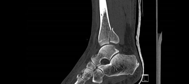
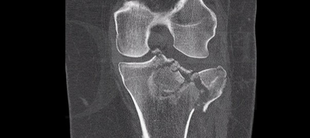
Tibiakopf-Verletzungen wurden von Schatzker auf der Grundlage von Röntgenaufnahmen in 6 Typen eingeteilt. Typen 5 (bikondylär) und Typ 6 (bikondylär mit vollständiger Trennung der Gelenkfläche von der Tibiaschafthülle) stellen die hochenergetischen Verletzungen dar. Eine neuere Klassifikation von Luo, basierend auf CT-Scans, unterteilt den Tibiakopf in 3 Säulen, medial, lateral und posterior, und hilft, die chirurgische Vorgehensweise bei der Fraktur je nach genauer Frakturkonfiguration zu leiten.

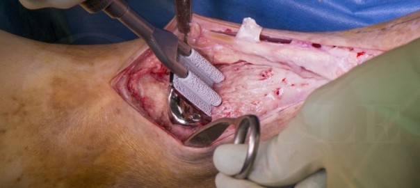
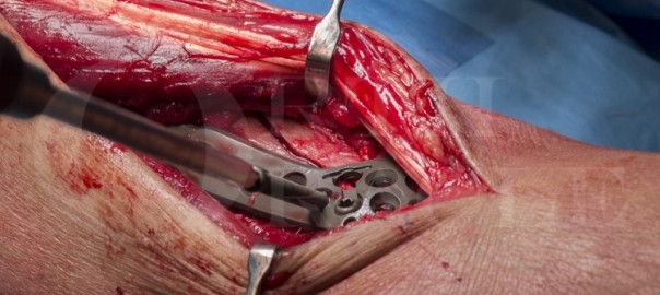
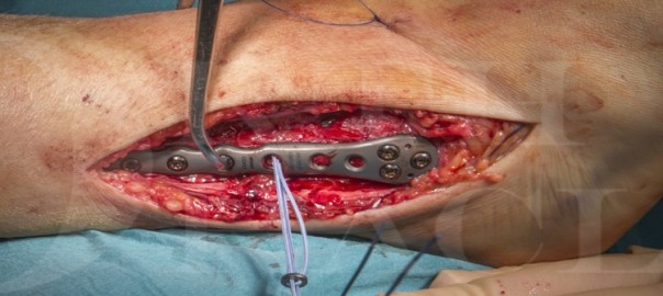
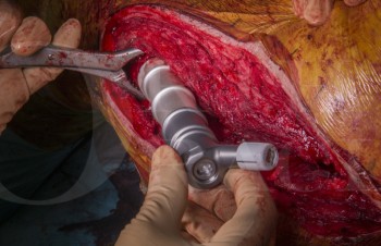
Die Mehrheit der Tibiakopf-Frakturen betrifft die laterale Seite und kann mit dem Standard anterolateralen Zugang mit Meniskusanhebung angegangen werden. Für Frakturen mit medialer oder posteriorer Beteiligung sind weitere Zugänge möglich, darunter direkter medialer, postero-medialer, direkter posteriorer oder sogar posterolateraler Zugang. Die Fixierung bikondylärer Tibiakopf-Frakturen über einen einzelnen anterioren Mittellinien-Schnitt wird heutzutage im Allgemeinen vermieden, da hierfür eine umfangreiche Weichteildissektion erforderlich ist und die damit verbundenen Wundprobleme.

Original Intro:

Tibial plateau fracture: 360 degree approach, anterior approach(stage 2), variable angle proximal tibia plate (DePuy-Synthes)

Learn the Tibial plateau fracture: 360 degree approach, anterior approach(stage 2), variable angle proximal tibia plate (DePuy-Synthes) surgical technique with step by step instructions on OrthOracle. Our e-learning platform contains high resolution images and a certified CME of the Tibial plateau fracture: 360 degree approach, anterior approach(stage 2), variable angle proximal tibia plate (DePuy-Synthes) surgical procedure.

As with much of adult trauma tibial plateau fractures have a bimodal distribution. In younger patients these injuries result from high energy mechanisms such as falls from height or road traffic accidents whereas in the elderly population they more often result from simple falls.

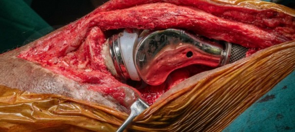
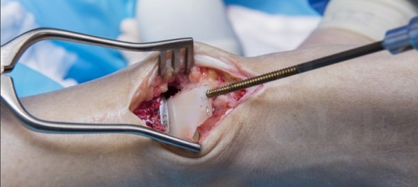
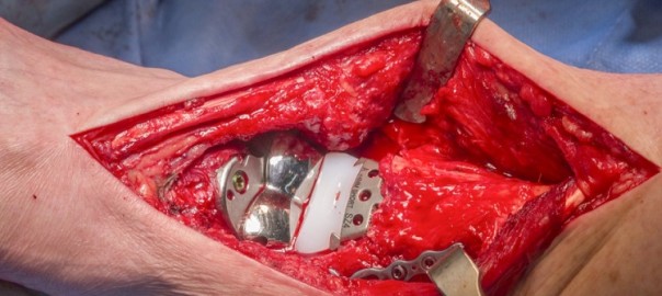
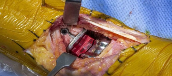
Tibial plateau injuries have been classified by Schatzker, on the basis of plain film radiographs, into 6 types. Types 5 (bicondylar) and type 6 (bicondylar with complete separation of the joint surface from the tibial shaft) represent the higher energy injuries.  A more recent classification by Luo, based on CT scans, divides the tibial plateau into 3 columns, medial, lateral and posterior and helps to guide the surgical approach to the fracture depending on the precise fracture configuration.

The majority of tibial plateau fractures involve the lateral side and can be approached with the standard anterolateral approach with meniscal elevation.  For those fractures with medial or posterior involvement a number of further approaches are possible from direct medial, postero-medial, direct posterior or even poster-lateral.  Fixation of bicondylar tibial plateau fractures via a single anterior mid-line incision is now generally avoided owing to extensive soft tissue dissection required and the consequential wound problems associated with this approach.

This technique should be read in conjunction with Tibial plateau fracture: 360 degree approach (stage 1).

Readers will find the following OrthOracle instruction techniques also of interest:

Tibial plateau fracture: 360 degree approach, posterior approach(Stage 1), using variable angle LCP proximal tibial plate (DePuy-Synthes).

Tibial plateau fracture: Posterior column fracture fixed with Synthes proximal tibial LISS plate
Tibial plateau fracture: Fixation of a bicondylar fracture with a dual incision technique (Smith and Nephew EVOS plates)
Tibial Plateau Fracture: Sleeved MBT(DePuy) Total Knee Replacement

Author: Peter Biberthaler MD.

Institution: Technical University of Munich, Klinikum rechts der Isar, Munich, Germany.

Clinicians should seek clarification on whether any implant demonstrated is licensed for use in their own country.


Revision der totalen Knieendoprothese: Zweite Stufe unter Verwendung des Adler Pantheon-Ersatzes mit Rekonstruktion des Gastrocnemius-Lappens

Übersetzt aus dem Englischen:

Lernen Sie die chirurgische Technik zur Revision der totalen Knieendoprothese: Zweite Stufe unter Verwendung des Adler Pantheon-Ersatzes mit Rekonstruktion des Gastrocnemius-Lappens mit schrittweisen Anweisungen auf OrthOracle. Unsere E-Learning-Plattform enthält hochauflösende Bilder und eine zertifizierte Fortbildung (CME) des chirurgischen Verfahrens zur Revision der totalen Knieendoprothese: Zweite Stufe unter Verwendung des Adler Pantheon-Ersatzes mit Rekonstruktion des Gastrocnemius-Lappens.

Die Anzahl der primären und revidierten Knieendoprothesen steigt stetig, was zwangsläufig zu mehr Protheseninfektionen führt, die von spezialisierten multidisziplinären Teams für Protheseninfektionen behandelt werden. Infektionen sind eine verheerende Komplikation der totalen Gelenkendoprothetik und nicht nur die häufigste Ursache für frühzeitiges Versagen von Gelenkersatzoperationen, sondern auch die häufigste Ursache für das Versagen von revisionsbedürftigen Knieendoprothesen zu jedem Zeitpunkt.

Dass Protheseninfektionen mit einer höheren Sterblichkeitsrate verbunden sind als einige häufige Krebserkrankungen, wurde weit verbreitet berichtet. Die Fünfjahresüberlebensrate nach einer Protheseninfektion beträgt 78% im Vergleich zu 90% bei Patienten, die sich einer aseptischen Revisionsendoprothetik unterziehen (Matar H, et al. Septic Revision Total Knee Arthroplasty Is Associated With Significantly Higher Mortality Than Aseptic Revisions: Long-Term Single-Center Study (1254 Patients). Journal of Arthroplasty 2021).

Original Intro:

Revision total knee replacement: Second stage using Adler Pantheon replacement with gastrocnemius flap reconstruction

Learn the Revision total knee replacement: Second stage using Adler Pantheon replacement with gastrocnemius flap reconstruction surgical technique with step by step instructions on OrthOracle. Our e-learning platform contains high resolution images and a certified CME of the Revision total knee replacement: Second stage using Adler Pantheon replacement with gastrocnemius flap reconstruction surgical procedure.

Increasing numbers of primary and revision knee replacements are being performed which inevitably lead to more prosthetic joint infections (PJI) presenting to specialist PJI multi-disciplinary teams. Infection is a devastating complication of total joint arthroplasty and not only the most common cause for early failure of joint replacements but also the most common cause for failure of revision knee replacements at any time.

That PJI is associated with higher mortality than some common malignancies has been widely reported. The five year survival after PJI is 78% compared to 90% in patients undergoing aseptic revision arthroplasty (Matar H, et al. Septic Revision Total Knee Arthroplasty Is Associated With Significantly Higher Mortality Than Aseptic Revisions: Long-Term Single-Center Study (1254 Patients). Journal of Arthroplasty 2021).

The treatment of prosthetic joint infection typically requires surgery involving explant of the infected prosthesis, radical debridement and then either immediate reimplantation or use of an antibiotic loaded cement spacer and delayed reimplantation  (a two-stage revision, as in this case). Alternative strategies include debridement and implant retention with modular exchange (indicated in acute PJI) and single-stage revision (considered in infected primary arthroplasty implants, sensitive organisms and without soft-tissue defects requiring plastic surgery). There is endless debate about selecting the correct option for each case, the decision is multi-factorial and probably best decided in specialist PJI multi-disciplinary meetings.

Infected revision knee replacements are increasing in prevalence due to the increasing numbers of revision joint replacements being performed for septic and aseptic indications such that a 7.5-fold increase in re-revision knee replacements due to infection that has been experienced in the UK since 2005 (Lenguerrand et al. Description of the rates, trends and surgical burden associated with revision for prosthetic joint infection following primary and revision knee replacements in England and Wales: an analysis of the National Joint Registry for England, Wales, Northern Ireland and the Isle of Man. 

Here I present a challenging case of reconstruction of an infected revision knee replacement, which underwent a two-stage revision using a porous tibial cone for severe bone loss and a silver-coated endoprosthetic replacement to manage segmental bone loss.

Readers will also find the following related OrthOracle surgical techniques of interest:

First Stage Revision Total Knee Replacement for Acute Prosthetic Joint Infection (Zimmer Biomet articulating spacer)

Second Stage Revision Total Knee Replacement. PFC / MBT TKA with metaphyseal sleeve and stem (Depuy)

Revision total Knee Replacement: Legion CCK (Smith and Nephew)

Revision total Knee Replacement- Legion Rotating Hinge Knee ( Smith and Nephew)

Revision Total Knee replacement (Tibia only): Smith and Nephew Legion revision knee system and Implantcast Epore tibial cone

First stage re-revision total knee replacement of Implantcast MUTARS MK EPR using reinforced static antibiotic cement spacer

Revision total Knee replacement: Second-stage with distal femoral EPR (Implantcast MUTARS MK) and EPORE collar and tibial cone

Revision Total Knee Replacement: Second stage using PFC / MBT with metaphyseal sleeve and stem (Depuy)

Total knee replacement: First stage revision with a hinged spacer and tibial tubercle osteotomy


Author: Jonathan Stevenson FRCS (Tr & Orth)

Institution: The Royal Orthopaedic Hospital, Birmingham, UK

Clinicians should seek clarification on whether any implant demonstrated is licensed for use in their own country.


Schulungen Datenschutz

Lösungen für alle Datenschutzfragen. Externer Datenschutzbeauftragter, Datenschutzpaket und Schulungen mit 20% Preisvorteil für BVOU-Mitglieder in der Akademie Deutscher Orthopäden.

Vorteile auf einen Blick

  • Seien Sie auf der sicheren Seite: externer Datenschutzbeauftragter plus Datenschutz-Check-up und Erstellung eines Datenschutzkonzepts
  • Unterstützung Ihrer schon bestehenden Datenschutzstrukturen mit unserem Datenschutzpaket
  • Datenschutzschulungen für niedergelassene Ärzte und deren Angestellte
  • E-Learning mit der meduplus Smart Learning® Methode mit nachweisbarem Lernerfolg
  • Online und mobil – Auf allen Endgeräten und ohne Softwareinstallation sofort starten!

Kursangebot

  1. Kurs „Schweigepflicht, Datenschutz und Kommunikation“
  2. Kurs „Cybersecurity und Datenschutz“
  3. Paket „Cybersecurity und Datenschutz für das Praxisteam“

Sprunggelenkersatz – STAR-Sprunggelenkersatz (Revision des Meniskuskomponents)

Übersetzt aus dem Englischen:

Lernen Sie die chirurgische Technik des Sprunggelenkersatzes – STAR-Sprunggelenkersatz (Revision des Meniskuskomponents) mit schrittweisen Anweisungen auf OrthOracle. Unsere E-Learning-Plattform enthält hochauflösende Bilder und eine zertifizierte Fortbildung (CME) des chirurgischen Verfahrens des Sprunggelenkersatzes – STAR-Sprunggelenkersatz (Revision des Meniskuskomponents).

Das Polyethylenlager eines mobil gelagerten Sprunggelenkersatzes kann brechen, verschleißen oder sich verschieben.

Wenn dies geschieht, muss der gesamte Sprunggelenkersatz möglicherweise überarbeitet werden, oder nur das meniskale Lager – unter Berücksichtigung der Tatsache, dass möglicherweise auch Verschleiß an den Metallflächen der Talus- und Tibiakomponenten aufgetreten ist.

Es ist wichtig, jegliche auslösenden Faktoren zu identifizieren, die zum Versagen des Meniskus geführt haben könnten (wie Sprunggelenkinstabilität oder Komponentenfehlstellung) und diese gleichzeitig mit der Überarbeitung des Lagers zu korrigieren.

Für diejenigen, die eine Sprunggelenkersatzoperation durchführen, sollte die Fähigkeit, ihre potenziellen Komplikationen (sowohl kurz- als auch langfristig) zu identifizieren und zu bewältigen, in Betracht gezogen werden.

Die folgende Operation zeigt meine Technik zur Bewältigung des isolierten Versagens einer meniskalen Komponente eines STAR-Sprunggelenkersatzes, einer der Implantate mit der längsten Erfolgsgeschichte.

Original Intro:

Ankle replacement-Star ankle replacement (revision of mensical component)

Learn the Ankle replacement-Star ankle replacement (revision of mensical component) surgical technique with step by step instructions on OrthOracle. Our e-learning platform contains high resolution images and a certified CME of the Ankle replacement-Star ankle replacement (revision of mensical component) surgical procedure.

The polyethylene bearing of a mobile bearing ankle replacement may fracture, wear or dislocate.

If this happens, the whole ankle replacement may need to be revised, or just the meniscal bearing – mindful of the fact that some wear may also have occurred to the metal surfaces of talar and tibial components.

It is important to identify any precipitating factors which may have caused the meniscus to fail (such as ankle instability or component malalignment) and to correct these at the same time as revising the bearing.

For those undertaking ankle replacement surgery the ability to identify and deal with its potential complications (both in the short and longer  term ) should be a consideration.

The following operation demonstrates my technique for managing the isolated failure of a meniscal component of a STAR ankle replacement , one of the implants with the longest track record.

Author: Paul Cooke FRCS 

Institution :The Nuffield Orthopaedic centre , Oxford ,UK.

Clinicians should seek clarification on whether any implant demonstrated is licensed for use in their own country.

zum Artikel auf OrthOracle

zum AOUC-Angebot für OrthOracle

Sprunggelenkersatz – Revision mit dem Wright Invision Sprunggelenkersatzsystem

Übersetzt aus dem Englischen:

Lernen Sie die chirurgische Technik der Sprunggelenkersatz-Revision mit dem Wright Invision Sprunggelenkersatzsystem mit schrittweisen Anweisungen auf OrthOracle. Unsere E-Learning-Plattform enthält hochauflösende Bilder und eine zertifizierte Fortbildung (CME) des chirurgischen Verfahrens der Sprunggelenkersatz-Revision mit dem Wright Invision Sprunggelenkersatzsystem.

Mit der zunehmenden Anzahl von Operationen zur totalen Sprunggelenkendoprothetik (TAA) weltweit ist zu erwarten, dass wir häufiger und wahrscheinlich komplexere Fälle von Versagenssituationen erleben werden. Das Versagen von Sprunggelenkersatzimplantaten tritt aufgrund verschiedener Ursachen auf, und einige Risiken für ein frühzeitiges Versagen wurden in der Literatur identifiziert, Gadd et al. Im Laufe der Zeit kann davon ausgegangen werden, dass Abnutzung eine Zytokinreaktion und Knochenlyse hervorruft, was zu Implantatmigration und Zystenbildung führt. Dieser Versagensmodus wird häufiger bei bestimmten Typen von TAA beobachtet, wobei das bekannteste Beispiel das mittlerweile zurückgezogene AES-System ist, das in einigen Serien massive Osteolyse verursachte.

Die Ursprünge von Zystenbildung und Lockerung wurden ausgiebig diskutiert. Rodriguez et al. (2010) hypothetisierten, dass die Zystenbildung mit der Pumpaktion zusammenhängen könnte, die zu synovialer Einschließung führt. Bonnin et al. (2011) hingegen waren der Meinung, dass einige der Zysten aus bereits vorhandenen arthritischen Zysten entstanden sein könnten. Jacobs et al. (2006) vermuteten, dass die Zystenbildung mit einer Überlastung eines lokalen afferenten Transportsystems mit Abriebpartikeln zusammenhängen könnte, was zu einer Ansammlung von Abriebpartikeln im periprothetischen Gewebe führt. Schließlich konnten einige Autoren in Rückgewinnungsproben aus den Zysten keine signifikanten Abriebpartikel finden, aber einige (Koivu et al.) stellten fest, dass es zu einer Aktivierung einer entzündlichen Kaskade kommt und schlossen daraus, dass es eine erhöhte Expression des Rezeptors für fortgeschrittene Glykierungsendprodukte und anderer “Gefahr-Signale” gibt, die zur Entzündung um die Implantate, zur Bildung multilokulärer Zysten und zur Osteolyse bei fehlgeschlagenen TAA-Implantaten beitragen könnten.

Original Intro:

Ankle replacement-Revision using Wright Invision Ankle replacement system

Learn the Ankle replacement-Revision using Wright Invision Ankle replacement system surgical technique with step by step instructions on OrthOracle. Our e-learning platform contains high resolution images and a certified CME of the Ankle replacement-Revision using Wright Invision Ankle replacement system surgical procedure.

With increasing numbers of total ankle arthroplasty (TAA) operations being carried out worldwide it can be expected that we are going to encounter more frequent and probably more complex failure situations. Failure of ankle replacements occurs due to a variety of causes and some risks for early failure have been established in the literature, Gadd et al. Over time it can be considered that wear will generate a cytokine response and bone lysis, leading to implant migration and cyst formation. This mode of failure is more frequently seen in certain makes of TAA with the best know example being the now withdrawn AES system which generated massive osteolysis in some series .

The origin of cysts and loosening has been widely debated. Rodriguez et al. () hypothesized that cyst formation may be related to pump action leading to synovial inclusion. However Bonnin et al. () felt that some of the cysts may have evolved from pre-existing arthritic cysts. Jacobs et al. () hypothesized that cyst formation may be related to overwhelming of a local afferent transport mechanism with wear particles, resulting in an accumulation of wear particles in periprosthetic tissue. Finally repeatedly authors have failed to find significant wear particles in retrieval specimens from the cysts but some (Koivu et al) have found that there is a switching on of an inflammatory cascade concluding that there is an increased expression of ‘high-mobility group box 1’ receptor for advanced glycation end product and other ‘danger signals’ which could contribute to inflammation around the implants, multilocular cyst formation, and osteolysis in failed TAA implants

The Mobility TAA (DePuy) was launched in 2003 and was  withdrawn in June 2014 . There have been no widespread publications of osteolysis though persistent medial pain is reported in some patients.

For failed TAA the commonest option is to convert the arthroplasty to an arthrodesis, which is an accepted and successful technique. Bone graft is often used from the fibula, iliac crest, allograft or using bone substitutes  to fill the resulting bone void from where the implant has been removed. If union occurs then pain relief will usually follow, but so does stiffness. If the bone loss is significant then the subtalar joint will often also be sacrificed with even greater loss of motion and functional deficit. Preservation of the subtalar joint is technically possible, usually requiring grafting of the talar defect and plate fixation of the ankle joint, if enough talar bone stock remains.

The alternative, in order to preserve motion, is to convert the failed arthroplasty to a revision joint replacement. Specific equipment has now come on to the market to facilitate this. The most tried and tested of these is the InBone system from Wright Medical Technologies, USA. There are publications that support its efficacy at early follow up (Devries, et al). The InBone is a fixed point, jig driven system using image intensification throughout in order to position the revision replacement in a reliable orientation. The system has been added to in the InVision System which also gives metallic solutions to bone loss by use of bulkier tibial trays and talar domes and with the addition of a flat plate to the talus, allowing bridging of cyst defects in the talus. The plate has augments which can be added to the plantar talar surface, though they are limited by their position.

Detailed is a complex  revision of a Mobile bearing De Puy Mobility Total Ankle Arthroplasty (which failed due to cyst formation) revised to a fixed bearing InVision Ankle replacement. The case is complicated by bony deficiency of the talus and fracture of the medial malleolus both of which are treated and discussed.

Gadd RJ, Barwick TW, Paling E, Davies MB, Blundell CM. Assessment of a three-grade classification of complications in total ankle replacement. Foot Ankle Int. 2014;35(5):434-437.

Kokkonen A, Ikavalko M, Tiihonen R, et al. High rate of osteolytic lesions in medium-term followup after the AES total ankle replacement. Foot Ankle Int. 2011 Feb;32(2):168-75.

Author : Mr Chris Blundell FRCS (Tr & Orth)

Institution :The Northern general hospital ,Sheffield ,UK.

Clinicians should seek clarification on whether any implant demonstrated is licensed for use in their own country.

Artikel auf OrthOracle

zum AOUC-Angebot für OrthOracle

Sprunggelenkersatz: Salto Totalgelenkersatz (Integra)

Übersetzt aus dem Englischen:

Lernen Sie die chirurgische Technik des Sprunggelenkersatzes: Salto Totalgelenkersatz (Integra) mit schrittweisen Anweisungen auf OrthOracle. Unsere E-Learning-Plattform enthält hochauflösende Bilder und eine zertifizierte Fortbildung (CME) des chirurgischen Verfahrens des Sprunggelenkersatzes: Salto Totalgelenkersatz (Integra).

Die totale Sprunggelenkendoprothetik (TAA) wird immer häufiger angewendet und ist eine zuverlässige Alternative zur Sprunggelenksfusion bei fortgeschrittener Sprunggelenksarthrose. In Großbritannien sind die Altersgruppen ähnlich wie bei anderen Endoprothesen – sie werden routinemäßig bei Personen über 60 Jahren durchgeführt und bei jüngeren Patienten nur unter bestimmten Umständen.

Es gibt viele neue Arten von Sprunggelenkersatzimplantaten auf dem Markt, und dies ist der größte Wachstumsbereich in der Herstellung von Endoprothesen. Allerdings haben nur wenige Implantate Ergebnisse über einen Zeitraum von mehr als 5 Jahren veröffentlicht, und der Maßstab, der zur Bewertung der Endoprothetik der unteren Extremität verwendet wird, beträgt in der Regel 10 Jahre. Daher ist es wichtig, den Typ des Sprunggelenkersatzes sorgfältig auszuwählen. Von den derzeit veröffentlichten Langzeitstudien entspricht die Überlebensrate der besseren Implantate derjenigen von Knieendoprothesen über diesen Zeitraum.

Der Ersatz ähnelt sehr dem einer totalen Knieendoprothese, mit modularen (dreiteiligen) oder festen (zweiteiligen) Lagerungsoptionen. Die gekrümmte Metallkuppel des Talusimplantats und der flache Plafond des Tibiaimplantats werden durch einen Einsatz aus ultrahochmolekularem Polyethylen getrennt, der auf jeder seiner Oberflächen diesen beiden verschiedenen Geometrien entspricht.

Original Intro:

Ankle Replacement: Salto Total Ankle replacement (Integra)

Learn the Ankle Replacement: Salto Total Ankle replacement (Integra) surgical technique with step by step instructions on OrthOracle. Our e-learning platform contains high resolution images and a certified CME of the Ankle Replacement: Salto Total Ankle replacement (Integra) surgical procedure.

Total ankle arthroplasty (TAA) is becoming more common and is a reliable alternative to ankle fusion, for end stage ankle arthritis.  In the UK, the age groups are similar to that of other arthroplasties – being routinely performed in the over 60’s and, in younger patients only under certain circumstances.

There are many new types of ankle replacement on the market and it is the largest growth area in arthroplasty manufacture. However only a few implants have published results over 5 years, and the benchmark used to judge lower limb arthroplasty is more commonly 10 years   Therefore, it’s important to select the type of ankle replacement carefully.  Of the longer term studies currently in publication, the survivorship of the better implants matches that of knee replacements over this time frame.

The replacement is very similar to a total knee replacement, with modular (3 part) or fixed bearing (2 part) options.  The curved metal dome of the talar implant and the flat plafond of the tibial implant are separated by an ultra-high molecular weight polyethylene insert, that conforms on each of its surfaces to these two different geometries.

The TAA is inserted through an anterior approach in nearly all cases and there are a wide variety of techniques and jigs for preparing the joint, with no one system unequivocally superior.

An ankle replacement is a complex procedure,  performed in a restricted anatomical region.  There is only a small margin for error with bone resection as well as a poorer wound healing environment than either a hip or knee replacement. There is a long learning curve and higher complication rates, than with hip or knee replacements with the volume of suitable cases being low – approximately 1% of the numbers of knee replacements being performed in the UK.

The functional outcomes, when comparing ankle replacement and ankle fusion, show understandable improvements in the range of movement but little difference in function or levels of pain.

Previously, the surgical complications were thought to be much higher in ankle arthroplasty, but recent studies shown that the complication rates are now very similar when compared to open arthrodesis.

As with all arthroplasties, the longevity and wear of the implant needs to be considered carefully, especially in younger patients. Current evidence though shows high success rates of conversion of a failed ankle replacement to a fusion, thereby alleviating some of this concern.

The decision whether to perform an ankle fusion or ankle replacement remains very individualised, depending on the experience of the surgeon and multiple patient factors, including age and especially the health and vascularity of the local soft tissues. Certainly, pre-existing arthritis in the surrounding hindfoot joints is a potential contra-indication to isolated ankle arthrodesis and a solid indication for ankle arthroplasty.

The Salto total ankle replacement has a 17 year track record, and is an implant that I have found reliable with jigging that aids reproducible implantation.   Specific features that differentiate it are various:
An anatomical talar design with differing medial and lateral curvatures – this replicates the anatomical curvature of the normal talus (many other implants don’t have this anatomical design) and better tribology.
It is a 3 part modular design, where the meniscal insert is mobile and self-centering, which I believe positions it more accurately at the mechanical axis of the joint.  On first weightbearing, the implant should “self-centre”
Its implantation, although more complex than some other ankle replacements,  provides the ability to make multiple fine adjustments for length, translation and rotation of the jigs. This gives the surgeon optimal accuracy for joint positioning
The design of the integration surfaces also result in very good primary stability upon implantation, which further improves with osseo-integration.

OrthOracle readers will also find the following associated instructional techniques of interest:

Ankle replacement-Revision using Wright Invision Ankle replacement system

Ankle replacement-Wright Infinity ankle replacement

Ankle replacement-Wright Prophecy

Ankle replacement-Star ankle replacement (revision of mensical component)

Ankle Replacement-BOX total ankle replacement (MatOrtho)

Author: Pete Rosenfeld FRCS(Tr & Orth)

Institution: St Marys Hospital & The Fortius clinic, London, UK.

Clinicians should seek clarification on whether any implant demonstrated is licensed for use in their own country.

zum Artikel auf OrthOracle

zum AOUC-Angebot für OrthOracle

Sprunggelenkersatz – BOX Totalgelenkersatz des Sprunggelenks (MatOrtho)

Übersetzt aus dem Englischen:

Lernen Sie die chirurgische Technik des Sprunggelenkersatzes – BOX Totalgelenkersatz des Sprunggelenks (MatOrtho) mit schrittweisen Anweisungen auf OrthOracle. Unsere E-Learning-Plattform enthält hochauflösende Bilder und eine zertifizierte Fortbildung (CME) des chirurgischen Verfahrens des Sprunggelenkersatzes – BOX Totalgelenkersatz des Sprunggelenks (MatOrtho).

Der Sprunggelenkersatz steht seit den 1970er Jahren als Intervention bei Sprunggelenksarthrose zur Verfügung. Die ersten Implantate wurden mit der Annahme entwickelt, dass das menschliche Sprunggelenk als echtes Scharnier funktioniert. Sie wurden daher nur entworfen, um eine uniplanare Bewegung (plantar und dorsalflexion) zu ermöglichen und bestanden nur aus 2 mechanisch verbundenen Komponenten. Die implantierten Sprunggelenke funktionierten jedoch auch mit einem gewissen Maß an Rotation, die an der schwächsten Stelle im “Mechanismus” stattfinden musste. Angesichts der Robustheit der implantierten Sprunggelenkscharniere erwies sich dies als die Implantat-/Gelenkschnittstelle, was zwangsläufig zu einem frühen Implantatversagen führte.

Die nächste Generation von Sprunggelenkersatzimplantaten verwendete ein 3-Komponenten-Design, bei dem die tibialen und taliaren Komponenten durch einen UHMW-Polyethylen-Meniskus verbunden waren, der eine Rotation innerhalb des Gelenks selbst ermöglichte. Diese “beweglichen” Prothesen nutzten die Kongruenz der “artikulierenden” Oberflächen, um die eingeschränkten Kräfte zu reduzieren und die hohen Kontaktbelastungen zu überwinden, was zu einer Reduzierung des Polyethylenverschleißes und des mechanischen Lockerns der festen Komponenten führte. Diese anfänglichen Ersatzteile, deren Ergebnisse immer noch definieren, welche Lebensdauer ein Sprunggelenkersatz erreichen sollte, sind die STAR-, Beuchal-Pappas- und Salto-Implantate. Fortschritte wurden bei der Instrumentierung und Reproduzierbarkeit der Implantation erzielt. Im Allgemeinen sind ihre Überlebensraten nach 10 Jahren niedriger als die für Hüft- und Knieersatzimplantate gemeldeten. Die Überlebensraten des UK National Joint Registry liegen jetzt im Bereich von 80 Prozent.

Original Intro:

Ankle Replacement-BOX total ankle replacement (MatOrtho)

Learn the Ankle Replacement-BOX total ankle replacement (MatOrtho) surgical technique with step by step instructions on OrthOracle. Our e-learning platform contains high resolution images and a certified CME of the Ankle Replacement-BOX total ankle replacement (MatOrtho) surgical procedure.

Ankle replacement has been available as an intervention for ankle arthritis since the 1970s. The initial implants were engineered on the assumption that the human ankle joint functioned as a true hinge . They were therefore designed only to allow uniplanar movement (plantar and dorsiflexion) and comprised just 2 components which were mechanically linked. The ankle  joints they were implanted into however also functioned with a degree of rotation which had to occur at the weakest point in the “mechanism”. Given the robustness of the implanted ankle hinges this transpired to be the implant/joint interface which therefore led invariably to early implant failure.

The next generation of ankle replacements  used a 3 component design, in which the Tibial and Talar components were linked by a UHMW polyethylene meniscus which allowed rotation to occur within the joint itself. These ‘mobile-bearing’ prostheses used the congruity of the ‘articulating’ surfaces, to reduce the constrained forces and overcome the high contact stresses resulting in a reduction in polyethylene wear and mechanical loosening of the fixed components. These initial replacements whose results still define what longevity an ankle replacement should attain are the STAR , Beuchal-Pappas and Salto implants. Advancements have been made in the instrumentation and reproducibility of implantation. In general their 10 year survivorships are lower than reported for hip and knee replacements  UK National Joint Registry survival rates are now in the region of 80 percent.

Total ankle replacement is generally not be recommended for younger or higher demand patients due to concerns of  longevity due to accelerated wear of the implant, the exception being patients with severe poly-articular inflammatory arthropathy.

The BOX (Bologna-Oxford) ankle manufactured by MatOrth is a three component prosthesis. The Box ankle replacement prosthesis has been designed to maximise congruency throughout the arc of motion, aiming to mimic normal ankle biomechanics. The bearing surface of the tibial component has a subtle curve in the coronal plane to accommodate for varus/ valgus force through the talo-crural joint.  Biomechanical modelling has demonstrated both rolling and sliding motions take place at the talocrural joint. In theory, full congruence should reduce wear by avoiding edge-loading. Similar to the STAR prosthesis, two anchorage bars on the tibial platform of the BOX ankle replacement provide stable primary fixation to the tibial bone. A precisely cut talar component allows a good press fit of the talar component, which is further stabilised with two vertical pegs.

Primary stability of the Box ankle replacement components reduces micromotion assisting the circumstances  necessary to provide reliable bone ingrowth.

Readers will also find the following techniques of interest:

Ankle replacement-Revision using Wright Invision Ankle replacement system

Ankle replacement-Wright Infinity ankle replacement

Ankle replacement-Wright Prophecy

Ankle replacement-Star ankle replacement (revision of mensical component)

Ankle Replacement -De Puy Mobility

Author: Nick Cullen FRCS (Tr & Orth)

Institution: The Royal National Orthopaedic Hospital, Stanmore, London, UK.

Clinicians should seek clarification on whether any implant demonstrated is licensed for use in their own country.

zum Artikel auf OrthOracle

zum AOUC-Angebot für OrthOracle

Pilonfraktur: Interne Fixierung unter Verwendung einer Stryker AxSOS 3Ti-Platte

Übersetzt aus dem Englischen:

Lernen Sie die chirurgische Technik der Pilonfraktur: Interne Fixierung unter Verwendung einer Stryker AxSOS 3Ti-Platte mit schrittweisen Anweisungen auf OrthOracle. Unsere E-Learning-Plattform enthält hochauflösende Bilder und eine zertifizierte Fortbildung (CME) des chirurgischen Verfahrens der Pilonfraktur: Interne Fixierung unter Verwendung einer Stryker AxSOS 3Ti-Platte.

Pilonfrakturen sind Frakturen des distalen Tibiaplafonds und sind per Definition intraartikulär und in einem belasteten Gelenk, was sie zu schweren und oft lebensverändernden Frakturen macht. Die Fraktur entsteht durch eine Mischung aus Scher- und Druckbelastungen auf die distale Tibiametaphyse.

Pilonfrakturen machen zwischen 5 und 10 % aller Frakturen der unteren Extremität aus und sind aufgrund der beteiligten Energie mit einer hohen (15-55 %) Komplikationsrate verbunden. Eine erhebliche Rotationskraft kann auch distale Tibiafrakturen verursachen, die den Plafond einbeziehen, und dies sind ebenfalls Pilonfrakturen. Dieser Mechanismus hat jedoch normalerweise weniger schwere Schädigungen des Weichgewebes und weniger Kompromisse in Bezug auf die Gelenkfläche hinsichtlich Fragmentierung und Knorpelschäden.

Der häufigste Verletzungsmechanismus ist ein Sturz aus großer Höhe, und dieser Fall demonstriert dies, nachdem er durch einen 4 Meter hohen Sturz auf Beton durch ein schwaches Dach verursacht wurde.

Die große Debatte besteht darin, wann eine offene Repositionsinterne Fixierung (ORIF) oder eine minimalinvasive Plattenosteosynthese (MIPO) verwendet werden soll und wann ein externes Fixationsgestell (wie ein Ilizarov oder ein anderes Drahtkonstrukt) als definitive Behandlung eingesetzt werden soll. In meinen Händen und in einer Einheit mit ausgezeichneten Fertigkeiten in der Drahtfixation vor Ort behandeln wir in der Regel diejenigen Fälle mit sehr schweren Weichteilschäden oder bei denen die Gelenkfläche stark fragmentiert ist, mit einem Gestell. Die Fälle, bei denen die Grad der Gelenkfragmentierung weniger schwerwiegend ist, werden in der Regel mit Plattenfixierung behandelt, wie es bei dem hier vorgestellten Fall der Fall war.

Original Intro:

Pilon fracture: Internal fixation using Stryker AxSOS 3Ti plate

Learn the Pilon fracture: Internal fixation using Stryker AxSOS 3Ti plate surgical technique with step by step instructions on OrthOracle. Our e-learning platform contains high resolution images and a certified CME of the Pilon fracture: Internal fixation using Stryker AxSOS 3Ti plate surgical procedure.

Pilon fractures are fractures of the distal tibial plafond and by definition are intra-articular and in a load bearing joint, this renders them serious and often life changing fractures. The fracture is sustained by a mixture of shear and compressive loads to the distal tibial metaphysis.

Pilon fractures make up between 5 and 10% of all lower limb fractures and because of the energy involved are associated with a high (15 – 55%) complication rate. Significant rotational force can also cause distal tibial fractures which involve the plafond and these are also pilon fractures. This mechanism though usually has less severe soft tissue damage and less compromise to the articular surface in terms of comminution and cartilage damage.

The most frequent mechanism of injury is a fall from height and this case demonstrates this , being sustained after a 4 metre fall onto concrete through a weak roof .

The big debate is when to use ORIF or minimally invasive plate osteosynthesis (MIPO), and when to use an external fixation frame (such as an Ilizarov or other fine wire construct) as definitive treatment. In my hands and working in a unit with excellent fine wire fixation skills locally we tend to treat those cases where there is very severe soft tissue damage or where the articular surface is grossly comminuted with a frame. Those cases where the degree of articular comminution is less severe are usually treated with plate fixation as was the case presented here.

OrthOracle readers will also find the following instructional operative techniques of use:

Pilon fracture: C-type fixed using Smith and Nephew EVOS small fragment system.

Pilon Fracture: C-type fixed with Stryker AxSOS 3 Periarticular Plating System

Pilon fracture: Internal fixation using Stryker AxSOS 3Ti plate.

Ankle fracture: Arthrex tightrope for acute syndesmotic injury and Stryker Variax plate for fibula fracture

Ankle fracture : Fibula pro-tibia fixation technique with Stryker Variax plate.

Ankle fracture: Medial malleolar fixation with ASNIS screws

Ankle fracture: Postero-lateral plating of pronation-external rotation ankle fracture (posterior malleolus))

Ankle fracture: Lateral malleolar fixation using Acumed Fibula Rod System

Author: Mr Chris Blundell FRDS (Tr & Orth)

Institution: IThe Northern General Hospital, Sheffield, UK.

Clinicians should seek clarification on whether any implant demonstrated is licensed for use in their own country.

zum Artikel auf OrthOracle

zum AOUC-Angebot für OrthOracle

Sprunggelenkfraktur: Fixation des hinteren Malleolus unter Verwendung eines posteriomedialen Zugangs

Übersetzt aus dem Englischen:

Lernen Sie die chirurgische Technik der Sprunggelenkfraktur: Fixation des hinteren Malleolus unter Verwendung eines posteriomedialen Zugangs mit schrittweisen Anweisungen auf OrthOracle. Unsere E-Learning-Plattform enthält hochauflösende Bilder und eine zertifizierte Fortbildung (CME) des chirurgischen Verfahrens der Sprunggelenkfraktur: Fixation des hinteren Malleolus unter Verwendung eines posteriomedialen Zugangs.

Sprunggelenkfrakturen mit Beteiligung des hinteren Malleolus gelten als mit einem schlechteren Ergebnis verbunden, jedoch sind das Management und die Indikationen für die Fixierung des hinteren Malleolus noch unzureichend definiert. Die Fixierung von hinteren Malleolusfrakturen wurde erstmals von Lounsbury und Metz im Jahr 1922 beschrieben, die einen extensilen medialen Zugang verwendeten, um große hintere Fragmente mit einem Knochenstift zu fixieren. Die traditionelle Lehre empfahl anschließend die Fixierung von hinteren Malleolusfragmenten basierend auf ihrer Größe, wenn sie auf einem seitlichen Röntgenbild beurteilt wurden, wobei die Fixierung für Fragmente empfohlen wurde, die ein Viertel oder ein Drittel der Gelenkfläche darstellen.

In letzter Zeit hat es einen Paradigmenwechsel in der Behandlung von hinteren Malleolusfrakturen gegeben, da erkannt wurde, dass die Größe des hinteren Malleolusfragments allein nicht der einzige Prädiktor für das Ergebnis ist. Faktoren wie Gelenkkongruenz, hintere Stabilität des Gelenks und Bandstabilität des Sprunggelenks sind ebenfalls wichtige Bestimmer des Ergebnisses.

Original Intro:

Ankle fracture: Posterior malleolar fixation using a posteromedial approach

Learn the Ankle fracture: Posterior malleolar fixation using a posteromedial approach surgical technique with step by step instructions on OrthOracle. Our e-learning platform contains high resolution images and a certified CME of the Ankle fracture: Posterior malleolar fixation using a posteromedial approach surgical procedure.

Ankle fractures with involvement of the posterior malleolus are recognised as having a poorer outcome however the management and indications for fixation of the posterior malleolus remain poorly defined. Fixation of posterior malleolar fractures was first described by Lounsbury and Metz in 1922 who used an extensile medial approach to fix large posterior fragments with a bone peg. Traditional teaching subsequently advocated fixation of posterior malleolar fragments based on their size when assessed on a lateral radiograph with fixation being recommended for fragments representing either a quarter or a third of the articular surface.

Recently there has been a paradigm shift in treatment of posterior malleolar fractures as it has become recognised that the size of the posterior malleolar fragment alone is not the only predictor of outcome. Factors such as articular congruence, posterior stability of the joint and ligamentous stability of the ankle are also important determinants of outcome.

There are a number of classifications describing the posterior malleolus however most are a development of that described by Haraguchi, dividing them into three types based on the appearance on axial CT.  The Haraguchi type 1 injury is a large posterolateral fragment involving the posterior syndesmotic ligament,   a Haraguchi type 2 posterior malleolar fracture extends medially and often has two distinct fragments: posterior and posteromedial with the posteromedial fragment extending to involve the posterior colliculus of the medial malleolus and thus the attachment of the deep deltoid can be rendered unstable by this fragment.  A type 3 injury is a sleeve avulsion of the posterior syndesmotic ligament, this pattern is usually not amenable to fixation and syndesmotic stability is achieved indirectly with syndesmotic screws or equivalent.

Surgical fixation of the posterior malleolus can be performed by a number of approaches.  Traditionally it was achieved with the patient in a supine position, indirect reduction of the fragment and fixation with screws inserted from anterior to posterior to capture the fragment.  It is increasingly recognised however that direct fixation of the fragment via a posterior approach is biomechanically superior.

The most common posterior approach is the posterolateral with an interval between the peronei and flexor hallucis longus, this affords good visualisation of the posterolateral fragment (Haraguchi 1) and also enables fixation of the fibula fracture via the same incision in most cases.  This approach however does not allow fixation of posterior malleolar fractures with medial extension and thus in my hands Haraguchi type 2 fractures are often better fixed via a posteromedial approach.  This can be performed in a variety of ways, often via the bed of tibialis posterior.  In this technique I describe an approach which allows fixation the posteromedial and posterolateral fragments via the same incision utilising windows either side of the posterior neurovascular bundle.

OrthOracle readers will also find the following associated instructional techniques of interest:

Ankle fracture: Postero-lateral plating of pronation-external rotation ankle fracture (posterior malleolus))

Posterior ankle decompression- Open technique

Tarsal tunnel decompression (for tarsal tunnel syndrome)

Ankle fracture: Medial malleolar fixation with ASNIS screws

Ankle fracture : Fibula pro-tibia fixation technique with Stryker Variax plate.

Ankle fracture: Lateral malleolar fixation using Acumed Fibula Rod System

Ankle fracture: Arthrex tightrope for acute syndesmotic injury and Stryker Variax plate for fibula fracture

Author: Paul Fenton, FRCS (Tr & Orth)

Institution: The Queen Elisabeth Hospital, Birmingham, UK.

Clinicians should seek clarification on whether any implant demonstrated is licensed for use in their own country.

zum Artikel auf OrthOracle

zum AOUC-Angebot für OrthOracle

Sprunggelenkfraktur: Arthrex TightRope für akute Syndesmosenverletzung und Stryker Variax-Platte für Fibulafraktur

Übersetzt aus dem Englischen:

Lernen Sie die chirurgische Technik der Sprunggelenkfraktur: Arthrex TightRope für akute Syndesmosenverletzung und Stryker Variax-Platte für Fibulafraktur mit schrittweisen Anweisungen auf OrthOracle. Unsere E-Learning-Plattform enthält hochauflösende Bilder und eine zertifizierte Fortbildung (CME) des chirurgischen Verfahrens der Sprunggelenkfraktur: Arthrex TightRope für akute Syndesmosenverletzung und Stryker Variax-Platte für Fibulafraktur.

Die distale Tibiofibulare Syndesmose ist ein einzigartiges syndesmotisches Gelenk, das durch eine Vielzahl von Bändern kraftvoll verbunden ist, um die Integrität des Sprunggelenks zu erhalten. Die distale Anatomie der beiden Knochen ist so gestaltet, dass die mediale Oberfläche der distalen Fibula in eine Rille auf der lateralen Oberfläche der distalen Tibia passt, die als Incisura fibularis bezeichnet wird und diesem Gelenk die knöcherne Stabilität bietet. Der vordere Teil dieses Gelenks wird durch das vordere untere distale Tibiofibulare Band (AITFL), der hintere Aspekt durch das hintere untere distale Tibiofibulare Band (PITFL) und das transversale Tibiofibulare Band stabilisiert. Direkt zwischen den benachbarten Oberflächen der Tibia und Fibula befindet sich das interosseöse Band, das sich über die gesamte Länge der Fibula erstreckt.

Das PITFL ist bei weitem das stärkste Band in diesem Komplex und am wenigsten wahrscheinlich zu reißen. Wenn es jedoch einmal gerissen ist, ist es am wahrscheinlichsten mit schweren Rotations- oder Luxationsverletzungen des Sprunggelenks und damit verbundener Sprunggelenkinstabilität verbunden.

Original Intro:

Ankle fracture: Arthrex tightrope for acute syndesmotic injury and Stryker Variax plate for fibula fracture

Learn the Ankle fracture: Arthrex tightrope for acute syndesmotic injury and Stryker Variax plate for fibula fracture surgical technique with step by step instructions on OrthOracle. Our e-learning platform contains high resolution images and a certified CME of the Ankle fracture: Arthrex tightrope for acute syndesmotic injury and Stryker Variax plate for fibula fracture surgical procedure.

The distal tibiofibular syndesmosis is a unique syndesmotic joint, powerfully bound by a variety of ligaments to maintain the integrity of the ankle mortise. The distal anatomy of the two bones are created in such a way that the medial surface of the distal fibula fits into a groove on the lateral surface of the distal tibia called the incisura fibularis which offers the bony stability to this joint. The anterior part of this joint is stabilised by the Anterior Inferior Distal Tibio-Fibular ligament (AITFL), the posterior aspect by the Posterior Inferior Distal Tibio-Fibular ligament (PITFL) and the transverse Tibio-Fibular ligament. Directly between the contiguous surfaces of the tibia and fibula is the interosseous ligament which extends throughout the length of the fibula.

The PITFL is by far the strongest ligament in this complex and is the least likely to be ruptured. Once ruptured however it is most likely to be associated with severe rotational or dislocating injuries of the ankle and associated ankle instability.

The syndesmotic ligaments  stabilise the fibula with respect to both talus and tibia, whilst allowing a degree of rotation of the fibula, required in particular during full ankle dorsiflexion when the widest portion of the talus comes into the mortise. As with any ligament injury the key point is not solely whether these ligaments have been injured, but rather their stability in determining the need for treatment. If instability is not addressed, high contact pressures at the joints surface ensue and early degenerative change occurs in most patients.

Traditional fixation of unstable syndesmotic injuries has been with two parallel, non-compressive, small fragment screws placed across the ankle at the level of the tibial incisura. Such fixation also requires a decision as to whether the screws need to be removed and furthermore,  the optimal timing for such removal.

The use of a Arthrex tightrope implant, is far more physiological in terms of the way the syndesmosis is held, allowing more normal movement at the ankle mortise. It is a simple and ingenious implant that has proved its worth not only in syndesmotic ankle reconstruction but also in the shoulder and forefoot. It consists of two metal buttons that rest on the respective bony  surfaces to be approximated, and a robust four stranded Fibrewire suture construct which links these buttons. The design allows easy apposition of the buttons towards each other by the effective “slip-knot” set up of the suture strands.

Readers will also find of use the following OrthOracle techniques:

Internal fixation of ankle fracture : Fibula pro-tibia fixation technique with Stryker Variax plate.

Internal fixation of medial malleolar ankle fracture with ASNIS screws

Lateral malleolar fixation using Acumed Fibula Rod System

Postero-lateral plating of pronation-external rotation ankle fracture (posterior malleolar fixation)

Fusion of the syndesmosis for isolated distal tibio-fibular arthritis

Author: Mark Herron FRCS

Institution: The Wellington Hospital, London, UK.

Clinicians should seek clarification on whether any implant demonstrated is licensed for use in their own country.

zum Artikel auf OrthOracle

zum AOUC-Angebot für OrthOracle