



Alle Beiträge von Lasse Walter

Teriparatid senkt das Risiko für Pseudarthrose und Revision bei Wirbelsäulenfusion

Artikel aus Zentralblatt für Orthopädie und Unfallchirurgie

In der spinalen Deformitätenchirurgie bei Erwachsenen besteht postoperativ ein hohes Risiko einer symptomatischen Pseudarthrose. Dieses Risiko ist bei Patienten mit Osteoporose doppelt so hoch im Vergleich zu Patienten mit normaler Knochendichte. Die vorliegende Studie analysiert den Einfluss von Teriparatid auf das Risiko für Pseudarthrosen und Revisionen nach operativer Versorgung adulter Wirbelsäulendeformitäten bei Patienten mit Osteoporose.

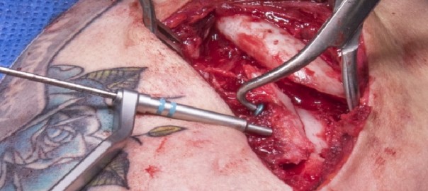
Schlüsselbeinbruch: Interne Fixation einer mehrfragmentären Fraktur mit der Stryker Variax 2 Verriegelungsplatte

Übersetzt aus dem Englischen:

Erlernen Sie die chirurgische Technik zur internen Fixation einer mehrfragmentären Schlüsselbeinfraktur mit der Stryker Variax 2 Verriegelungsplatte mit Schritt-für-Schritt-Anleitungen auf OrthOracle. Unsere E-Learning-Plattform enthält hochauflösende Bilder und eine zertifizierte CME des chirurgischen Verfahrens zur internen Fixation einer mehrfragmentären Schlüsselbeinfraktur mit der Stryker Variax 2 Verriegelungsplatte.

Schlüsselbeinfrakturen sind häufig und machen etwa 4% aller Frakturen aus, sowie bis zu 44% der Frakturen des Schultergürtels, wobei das mittlere Drittel bei weitem die häufigste Stelle ist. Die Behandlung solcher Verletzungen kann schwierig sein und das Ergebnis kann unbefriedigend sein. Es wurde eine Nicht-Verheilungsrate von 15 bis 20% bei dislozierten mittleren Drittel-Schlüsselbeinfrakturen berichtet. Diese kann durch chirurgische Intervention erheblich reduziert werden. Obwohl bei jeder chirurgischen Intervention Risiken und mögliche Komplikationen bestehen, zeigen veröffentlichte Ergebnisse, dass die Platten- und Schraubenfixation sicher durchgeführt werden kann, um ein gutes Ergebnis mit verbesserter Patientenzufriedenheit und einer Reduzierung der Nicht-Verheilungsrate im Vergleich zu konservativ behandelten Frakturen zu erzielen.

Original Intro:

Clavicle fracture: Internal fixation of comminuted fracture with Stryker Variax 2 locking plate

Learn the Clavicle fracture: Internal fixation of comminuted fracture with Stryker Variax 2 locking plate surgical technique with step by step instructions on OrthOracle. Our e-learning platform contains high resolution images and a certified CME of the Clavicle fracture: Internal fixation of comminuted fracture with Stryker Variax 2 locking plate surgical procedure.

Clavicle fractures are common accounting for around 4% of all fractures and up to 44% of fractures of the shoulder girdle of which the middle third is by far the most common site.  The management of such injuries can be difficult and outcome can be unsatisfactory.  There has been reported non-union rate of displaced mid-shaft clavicle fractures of between 15 and 20%.  This can be reduced significantly with surgical intervention.  Whilst there is risk and some potential complications with any surgical intervention the published results demonstrate that  plate and screw fixation can be performed safely to give a good outcome with improved patient satisfaction and a reduction in the non-union rate compared to conservatively managed fractures.

There are many implant companies with clavicle specific plates.  Described here is the use of the Stryker VariAx 2 clavicle locking plate system.  These contoured and sided plates are available for lateral injuries, superior shaft fractures and for use along the anterior face of the clavicle.  The plates and screws are made of different grades of titanium allowing the harder locking screws to cut their own thread within the softer material of the plate and their position can be adjusted up to three times.  Screws are available in sizes 3.5 and 2.7 in both non-locking and locked varieties.  The clavicle set also provides useful instruments to facilitate exposure with periosteal elevation, fracture reduction in terms of clamps and wires as well as plate holding clamps.

Readers will also find of interest the following OrthOracle instructional techniques:

Internal fixation of midshaft clavicle fracture using Synthes superior clavicle plate

Arthroscopic repair of acute Acromioclavicular Joint Separation with the Arthrex TightRope system

Author: Mark Crowther FRCS (Tr & Orth)

Institution: The Avon Orthopaedic centre, Bristol, UK.

zum Artikel auf OrthOracle

zum AOUC-Angebot für OrthOracle

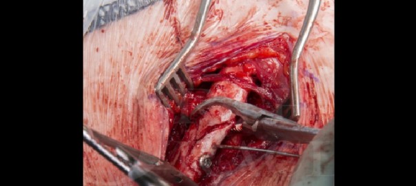
Schlüsselbeinbruch: Fixation mit der Synthes Superior Schlüsselbeinplatte

Übersetzt aus dem Englischen:

Erlernen Sie die chirurgische Technik zur Fixation eines Schlüsselbeinbruchs mit der Synthes Superior Schlüsselbeinplatte mit Schritt-für-Schritt-Anleitungen auf OrthOracle. Unsere E-Learning-Plattform enthält hochauflösende Bilder und eine zertifizierte CME des chirurgischen Verfahrens zur Fixation eines Schlüsselbeinbruchs mit der Synthes Superior Schlüsselbeinplatte.

Schlüsselbeinfrakturen sind häufig und machen etwa 2,6% aller Frakturen aus, wobei Frakturen im mittleren Drittel 80% ausmachen. 2-6% treten im medialen Drittel auf, während die verbleibenden 12-16% am lateralen Ende des Schlüsselbeins auftreten. Diese Frakturen kommen am häufigsten bei einer jungen und aktiven Bevölkerung vor.

Die Behandlung solcher Verletzungen kann umstritten sein, da dislozierte mittlere Drittel-Schlüsselbeinfrakturen historisch konservativ mit niedrigen gemeldeten Nicht-Verheilungsraten behandelt wurden. In der Literatur werden jedoch zunehmend deutlich höhere Raten von bis zu 20% berichtet. Es ist allgemein anerkannt, dass diese durch chirurgische Eingriffe erheblich reduziert werden können.

Obwohl bei jedem chirurgischen Eingriff ein gewisses Risiko besteht, zeigen veröffentlichte Ergebnisse auch, dass die Fixation mit Platten und Schrauben sicher durchgeführt werden kann, um ein gutes Ergebnis mit verbesserter Patientenzufriedenheit und einer Reduzierung der Nicht-Verheilungsrate im Vergleich zu konservativ behandelten Frakturen zu erzielen.

Original Intro:

Clavicle fracture: Fixation using Synthes superior clavicle plate

Learn the Clavicle fracture: Fixation using Synthes superior clavicle plate surgical technique with step by step instructions on OrthOracle. Our e-learning platform contains high resolution images and a certified CME of the Clavicle fracture: Fixation using Synthes superior clavicle plate surgical procedure.

Clavicle fractures are common accounting for around 2.6% of all fractures, with middle third fractures accounting for 80%. 2-6% occur in the medial third, while the remaining 12-16% occur in the lateral end of the clavicle. These fractures most commonly occur in a young and active population.

The management of such injuries can be controversial as historically, displaced mid-shaft clavicle fractures have been managed conservatively with low reported non-union rates. However, more literature has reported much higher rates, up to 20% on occasion. It is widely accepted that this can be reduced significantly with surgical intervention.

Whilst there is some risk with any surgical intervention, the published results also demonstrate that  plate and screw fixation can be performed safely to give a good outcome with improved patient satisfaction and a reduction in the non-union rate compared to conservatively managed fractures.

For displaced lateral end clavicle fractures, there is a higher rate of non-union and therefore, the threshold for surgical fixation is lower. Options for surgical fixation include specific lateral end anatomic clavicle plates utilising a variety of locking screw options for the smaller lateral fragment. Some plates allow reconstruction of the ruptured coracoclavicular ligaments with a non-absorbable material to augment and support the repair. If standard internal fixation is not possible due to comminution, these fractures can also be managed with a hook plate. The lateral end of this plate hooks under the acromion and butresses the lateral clavicle from displacing superiorly. The disadvantage with this type of fixation is that it needs to be removed once the fracture has healed to avoid impinging on the rotator cuff and causing a cuff tear. Whilst in situ, it may also physically limit the arc of movement in the shoulder.

There are many implant companies with clavicle specific plates. My preference is for the Synthes LCP clavicular (Locking Compression Plates). These are anatomically contoured and sided plates with either a superior or supero-anterior plates option for mid-shaft clavicle fractures. Superior clavicle plates tend to require less contouring. Theoretically, the medial screws are also directed further away from the neuro-vascular bundle. The plates and screws on the Synthes set are made of stainless steel with the “combi-hole” design allowing the option of either locking or non-locking screws within the same hole. The Synthes clavicle set also provides useful instruments to facilitate the surgical exposure and fracture reduction.

Author: Mr Sam Chan, FRCS (Tr & Orth).

Institution: The Queen Elizabeth hospital, Birmingham, UK.


zum Artikel auf OrthOracle

zum AOUC-Angebot für OrthOracle

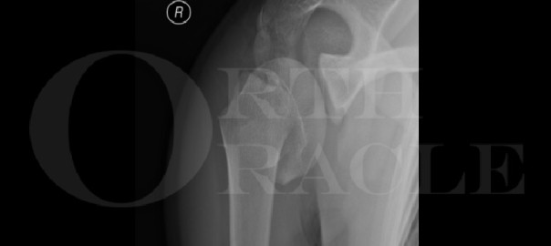
Humerusfraktur: Fixation einer proximalen Epiphysenfraktur mit der Synthes Philos Platte

Übersetzt aus dem Englischen:

Erlernen Sie die chirurgische Technik zur Fixation einer proximalen Epiphysenfraktur des Humerus mit der Synthes Philos Platte mit Schritt-für-Schritt-Anleitungen auf OrthOracle. Unsere E-Learning-Plattform enthält hochauflösende Bilder und eine zertifizierte CME des chirurgischen Verfahrens zur Fixation einer proximalen Epiphysenfraktur des Humerus mit der Synthes Philos Platte.

Proximale Humerusepiphysenfrakturen sind selten und machen weniger als 5% der Frakturen bei Kindern aus. Sie können grob in zwei Typen unterteilt werden: Salter-Harris I oder Salter-Harris II.

Eine Salter-Harris II ist die häufigere Verletzung und tritt typischerweise bei Jugendlichen nach einem stumpfen Trauma der Schulter auf.

Eine Salter-Harris I ist häufiger bei Kindern unter 5 Jahren. Salter-Harris I Verletzungen können als Überlastungsverletzung mit resultierender sekundärer Fraktur auftreten. Typischerweise kann eine Erweiterung der Epiphyse gesehen werden.

Die radiografische Auswertung der Schulter erfolgt mit Standard-AP- und axialen oder Skapula-Y-Ansichten der Schulter. Kontralaterale Schulteransichten können zum Vergleich angefordert werden.

Die kombinierten Ansichten ermöglichen eine Beurteilung des Schweregrads der Dislokation.

Original Intro:

Humeral fracture: Fixation of proximal physeal fracture using Synthes Philos plate

Learn the Humeral fracture: Fixation of proximal physeal fracture using Synthes Philos plate surgical technique with step by step instructions on OrthOracle. Our e-learning platform contains high resolution images and a certified CME of the Humeral fracture: Fixation of proximal physeal fracture using Synthes Philos plate surgical procedure.

Proximal humerus physeal fractures are uncommon and account for fewer than 5% of fractures in children. They can be broadly divided into 2 types ,Salter-Harris I or Salter-Harris II.

A Salter-Harris II is the more common injury and is typically an injury of adolescents following blunt trauma to the shoulder.

A Salter-Harris I is more common in children under 5 years of age. Salter-Harris I injuries can occur as an overuse injury with resultant secondary fracture. Typically, a widening of the physis can be seen.

Radiographic evaluation of the shoulder is performed with standard AP and axial or scapula Y views of the shoulder.
Contralateral shoulder views can be requested for comparison.

The combined views allow assessment of the severity of the displacement.

The epiphysis tends to abduct and externally rotate due to the pull of the rotator cuff (supraspinatus and infraspinatus). The humeral shaft is usually displaced into a shortened, adducted and anterior position due to the pull of deltoid and pectoralis major.

Anatomically, the proximal humeral physis contributes to 80% of humeral growth. It closes between the ages of 16-17 in females and at around 18 in males.

The decision making is for type of fixation is down to a balance of providing a stable reduction without compromising growth potential. Where growth arrest is not a concern, a Philos plate would provide the most robust fixation as it provides adequate fixation in the epiphyseal and metaphyseal fragments. In descending order of stability, this is followed by cannulated screws, flexible nails and Kirshner wire pinning.


Author: Mr Samuel Chan FRCS (Tr & Orth).

Institution: The Queen Elizabeth Hospital, Birmingham, UK.

zum Artikel auf OrthOracle

zum AOUC-Angebot für OrthOracle

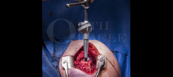
Totaler Schulterersatz: Lima SMR Reverse Shoulder für proximale Humerusfraktur

Übersetzt aus dem Englischen:

Lernen Sie den totalen Schulterersatz: Lima SMR Reverse Shoulder für proximale Humerusfraktur mit Schritt-für-Schritt-Anleitungen auf OrthOracle. Unsere E-Learning-Plattform enthält hochauflösende Bilder und eine zertifizierte CME des chirurgischen Verfahrens für den totalen Schulterersatz: Lima SMR Reverse Shoulder für proximale Humerusfraktur.

Es ist bekanntermaßen schwierig, bei älteren Patienten mit dislozierten, mehrfragmentären proximalen Humerusfrakturen, die chirurgisch behandelt werden, ein gutes funktionelles Ergebnis zu erzielen. In den letzten Jahren haben Schulterspezialisten erkannt, dass diese Verletzung als Fraktur mit Rotatorenmanschettendefizit behandelt werden sollte und daher vermehrt inverse total Schulterprothesen mit einer Prothesenkugel auf der Glenoidseite (der Glenosphäre) und einer Pfanne auf der Humerusseite eines halbkonstruierenden Systems verwenden. Dies medialisiert das Rotationszentrum und erhöht den Hebelarm, auf dem der große Deltamuskel arbeiten kann, in Abwesenheit der Rotatorenmanschettensehnen, die normalerweise um den proximalen Humerus eingesetzt sind. Das Hauptziel der inversen Schulterarthroplastik war es, das schwierige Problem der Arthritis in der Schulter mit Rotatorenmanschettendefizit, der sogenannten Rotatorenmanschettenarthropathie, zu lösen, die als chronischer Zustand auftritt. Solche Implantate sind in den letzten 30 Jahren weit verbreitet und haben an Popularität gewonnen, so dass sie die Anzahl der anatomischen Schulterersatzoperationen übertroffen haben.

In jüngerer Zeit werden die gleichen Implantate zur Behandlung von dislozierten mehrfragmentären Frakturen des proximalen Humerus eingesetzt. Traditionell sind solche Frakturen sowohl bei konservativer Behandlung als auch bei interner Fixation mit relativ schlechten Ergebnissen verbunden. Letztere wurde mit konturierten proximalen Humerusverriegelungsplatten erheblich erleichtert, dennoch ist eine solche Operation bei osteoporotischem Knochen technisch schwierig und oft mit schlechten Ergebnissen verbunden. Ein erhebliches Risiko besteht in der avaskulären Nekrose des Humeruskopfes und der Resorption der Tuberositäten mit daraus resultierender Rotatorenmanschettendysfunktion, die der Rotatorenmanschettenarthropathie ähnelt, weshalb die inverse Arthroplastik angewendet wird.

Eine Alternative wäre die Hemiarthroplastik-Rekonstruktion des proximalen Humerus unter Verwendung einer Hemiarthroplastikprothese mit Schaft und Rekonstruktion der Tuberositäten um das Implantat. In solchen Fällen besteht das signifikante Problem darin, dass die Tuberositäten resorbieren und der Patient mit einem Metallspacer und einer Schulter mit Rotatorenmanschettendefizit zurückbleibt.

Original Intro:

Total shoulder replacement: Lima SMR Reverse Shoulder for proximal humeral fracture

Learn the Total shoulder replacement: Lima SMR Reverse Shoulder for proximal humeral fracture surgical technique with step by step instructions on OrthOracle. Our e-learning platform contains high resolution images and a certified CME of the Total shoulder replacement: Lima SMR Reverse Shoulder for proximal humeral fracture surgical procedure.

It is notoriously difficult to achieve a good functional outcome in older patients with displaced comminuted proximal humerus fractures managed surgically.  In recent years shoulder surgeons have considered that this injury should be treated as a fracture with rotator cuff deficiency and hence have been using reverse geometry total shoulder replacements with a prosthetic ball on the glenoid (the glenosphere) and socket on the humeral side of a semi-constrained construct. This medialises the centre of rotation and increases the moment arm on which the large deltoid muscle can work in the absence of the rotator cuff tendons which normally insert around the proximal humerus. The main design remit for reverse shoulder arthroplasty was to address the difficult problem of arthritis in the rotator cuff deficient shoulder so called cuff tear arthropathy which presents as a chronic condition. Such implants have been in widespread use over the last 30 years gaining in popularity such that they have overtaken the number of anatomical shoulder replacements performed. 

More recently the same implants have been used to treat comminuted displaced fractures of the proximal humerus.  Traditionally such fractures have been associated with relatively poor outcomes when managed either conservatively or with internal fixation.  The latter has been made significantly easier with contoured proximal humeral locking plates but still such surgery in osteoporotic bone is technically difficult and often associated with poor outcomes.  A significant risk is avascular necrosis of the humeral head and resorption of the tuberosities with consequent rotator cuff dysfunction mimicking cuff tear arthropathy hence the adoption of reverse geometry arthroplasty.

An alternative would be to consider hemiarthroplasty reconstruction of the proximal humerus using a stemmed hemiarthroplasty prosthesis with reconstruction of the tuberosities around the implant.  In such cases the significant problem is that the tuberosities resorb and the patient is left with a metal spacer and a rotator cuff deficient shoulder. 

Implant companies have developed fracture specific reverse geometry implants which are tailored to the task. The Lima SMR is one of these as part of the spectrum of options available on their platform system. It uses the same glenosphere and humeral stem (cemented or uncemented) as their cuff tear arthropathy stemmed prosthesis but has a module trauma body added to the humeral stem which incorporates holes for passage of sutures to repair residual small fragments of bone and rotator cuff remnants.

Whilst there is some evidence in sporadic publications we are yet to have good hard level 1 evidence that reverse geometry arthroplasty in such cases is reliably and significantly better than either conservative management, internal fixation or hemiarthroplasty reconstruction, however there are trials ongoing to try and address the issue.  Anecdotally most shoulder surgeons will have many examples of good outcomes from reverse geometry arthroplasty in such cases.


Author: Mark Crowther FRCS (Tr & Orth)

Institution: The Avon Orthopaedic centre, Bristol, UK.

zum Artikel auf OrthOracle

zum AOUC-Angebot für OrthOracle

Totaler Ellenbogenersatz mit dem Coonrad-Morrey Total Elbow Replacement (Zimmer-Biomet)

Übersetzt aus dem Englischen:

Lernen Sie den totalen Ellenbogenersatz mit dem Coonrad-Morrey Total Elbow Replacement (Zimmer-Biomet) mit Schritt-für-Schritt-Anleitungen auf OrthOracle. Unsere E-Learning-Plattform enthält hochauflösende Bilder und eine zertifizierte CME des chirurgischen Verfahrens für den totalen Ellenbogenersatz mit dem Coonrad-Morrey Total Elbow Replacement (Zimmer-Biomet).

Ein Ellenbogengelenkersatz ist eine attraktive Möglichkeit, Schmerzen bei Patienten mit schmerzhaften arthritischen Gelenken zu lindern, insbesondere im Vergleich zu den Alternativen.

Die ersten Prothesen waren feste Scharniere (die nur Beugung und Streckung zuließen), die aufgrund der normalen Varus- und Valgusbewegungen der Ulna in Bezug auf den Humerus im Gelenk zu vorhersehbarem frühen Lösen der Komponenten führten. Unverbundene Komponenten (bei denen die Humerus- und Unterarmkomponenten nicht physisch verbunden sind und auf das Gleichgewicht der Weichteile für die Stabilität angewiesen sind) wurden entwickelt, aber diese neigten zur Luxation.

Original Intro:

Total elbow replacement using the Coonrad-Morrey Total Elbow Replacement (Zimmer-Biomet)

Learn the Total elbow replacement using the Coonrad-Morrey Total Elbow Replacement (Zimmer-Biomet) surgical technique with step by step instructions on OrthOracle. Our e-learning platform contains high resolution images and a certified CME of the Total elbow replacement using the Coonrad-Morrey Total Elbow Replacement (Zimmer-Biomet) surgical procedure.

An elbow replacement  is an attractive proposition to improve pain in patients that have painful arthritic joints, particularly when compared to the alternatives.

Initial prostheses were fixed hinges (allowing only flexion and extension) which, due to the normal varus and valgus toggle of the ulna in relation to the humerus at the joint, led to predictable early loosening of the components.  Unlinked components (with the humeral and the forearm components not being physically connected and relying on balancing the soft tissues for stability) were developed, but these were prone to dislocation.

Contemporary implants are therefore usually coupled (physically connected together), but with play in varus and valgus replicating the normal joint  to reduce the likelihood of mechanical loosening.  Nonetheless, loosening of the components (usually aseptic, but potentially secondary to infection) still frequently occurs, and therefore patients are recommended to restrict activities following elbow replacement for the remainder of their lives, in particular limiting lifting loads and torsion activities.  For this reason, the number of patients who will accept the required lifestyle restrictions needed for a replacement to so stand to benefit from elbow replacement after appropriate counselling are relatively limited.

Patients with inflammatory arthritis have generally been the main group of patients who underwent elbow replacement surgery, in large part as their functional demands have tended to be lower due to the involvement of multiple other joints with their disease.  With the advent of biologic treatments for systemic inflammatory arthropathies, the number of patients who need to consider having an elbow replacement has reduced. As the number of patients receiving elbow replacements for inflammatory arthritis has reduced, the proportion of patients receiving implantations for un-reconstructible trauma in the elderly, , has increased. The outcomes reported in this group has been good, at least in the medium term. The overall number of primary implantations entered into the UK National Joint Registry each year since data recording began (1st April 2012) has remained around 600 across England and Wales. Most patients having their elbow replaced are still those with an underlying inflammatory arthritis, ideally in their 60’s or older (to minimise the risk of revision surgery being necessary), but if younger patients understand the potential implications of a replacement and accept the restrictions still needed to maximise the longevity of the implant, elbow replacement can be offered to younger patients as well.

The Coonrad-Morrey total elbow replacement is the international market leading elbow replacement implant, used in its current form for around 20 years, with 10-year survivorship of 80-92% reported. In common with most elbow replacements, it is designed for cemented implantation. It represents a reliable, relatively straightforward component to insert with readily available exchange of the polyethylene bearings in the event of significant wear, in the presence of stable implants. The different component sizes have had interchangeable bearings since the early 2000’s to facilitate this.

Newer implants, such as the Latitude EV (Wright Medical), Discovery (Lima) and Nexel (Zimmer-Biomet), are available and are defining their roles in elbow replacement surgery. All have anterior flanges to the humeral component. The Latitude EV can be used as a hemiarthroplasty for treating distal humeral fractures, or as a total elbow replacement, and also has a radial head component. The Latitude also has the option of being inserted coupled (linked)or uncoupled (no linkage between the ulnar and humeral components, which may potentially reduce aseptic loosening), or can even be unlinked at a subsequent procedure once soft-tissue healing has provided stability (or, conversely, be coupled to restore stability if initially inserted unlinked).

The newer implants have advantages in certain technical areas such as easier independent insertion of the ulna and humeral components and where indicated, a radial head component. Whilst the ability to couple the implant once the cemented components are stable may have a theoretical advantages in terms of longevity, none of the newer implants have the data to support a comparable longevity to the Coonrad-Morrey implant. and the surgical techniques and instrumentation are a bit more involved and technically demanding.

I still use the Coonrad-Morrey for most primary replacements, in part due to familiarity, both my own and, as importantly, the theatre staffs. Total elbow replacement even in specialist centres is relatively infrequently performed, the scrub staff will have only a limited exposure to the procedure and the instrumentation. I think under such circumstances it is important to consistently use an appropriate implant, amongst a number of reasons so that the whole team become familiar with the steps and instruments to speed up and improve the operative procedures.

Until more contemporary implants demonstrate a definite survival advantage I will continue to use the Coonrad-Morrey for the majority of my patients, reserving use of a newer implants for younger patients and those in whom I think it is more likely that a revision will become necessary.


Author: Chris Little FRCS (Tr & Orth)

Institution: The Nuffield Orthopaedic centre, Oxford, UK.

zum Artikel auf OrthOracle

zum AOUC-Angebot für OrthOracle

Hintere Halswirbelsäulen-Laminoplastik (Bioplate-Kranialfixationssystem) und Entfernung eines C5-Nervenscheiden-Tumors

Übersetzt aus dem Englischen:

Lernen Sie die chirurgische Technik zur Tibiakopf-Fraktur: 360-Grad-Zugang, anteriore Vorgehensweise (Stufe 2), variable Winkel proximale Tibiaplatte (DePuy-Synthes) mit schrittweisen Anweisungen auf OrthOracle. Unsere E-Learning-Plattform enthält hochauflösende Bilder und eine zertifizierte Fortbildung (CME) des chirurgischen Verfahrens zur Tibiakopf-Fraktur: 360-Grad-Zugang, anteriore Vorgehensweise (Stufe 2), variable Winkel proximale Tibiaplatte (DePuy-Synthes).

Wie bei vielen Traumata im Erwachsenenalter haben Tibiakopf-Frakturen eine bimodale Verteilung. Bei jüngeren Patienten resultieren diese Verletzungen aus hochenergetischen Mechanismen wie Stürzen aus großer Höhe oder Verkehrsunfällen, während sie in der älteren Bevölkerung häufiger durch einfache Stürze verursacht werden.

Tibiakopf-Verletzungen wurden von Schatzker auf der Grundlage von Röntgenaufnahmen in 6 Typen eingeteilt. Typen 5 (bikondylär) und Typ 6 (bikondylär mit vollständiger Trennung der Gelenkfläche von der Tibiaschafthülle) stellen die hochenergetischen Verletzungen dar. Eine neuere Klassifikation von Luo, basierend auf CT-Scans, unterteilt den Tibiakopf in 3 Säulen, medial, lateral und posterior, und hilft, die chirurgische Vorgehensweise bei der Fraktur je nach genauer Frakturkonfiguration zu leiten.

Die Mehrheit der Tibiakopf-Frakturen betrifft die laterale Seite und kann mit dem Standard anterolateralen Zugang mit Meniskusanhebung angegangen werden. Für Frakturen mit medialer oder posteriorer Beteiligung sind weitere Zugänge möglich, darunter direkter medialer, postero-medialer, direkter posteriorer oder sogar posterolateraler Zugang. Die Fixierung bikondylärer Tibiakopf-Frakturen über einen einzelnen anterioren Mittellinien-Schnitt wird heutzutage im Allgemeinen vermieden, da hierfür eine umfangreiche Weichteildissektion erforderlich ist und die damit verbundenen Wundprobleme.

Original Intro:

Posterior Cervical Laminoplasty (Bioplate cranial fixation system) and Excision Of C5 nerve sheath tumour

Learn the Posterior Cervical Laminoplasty (Bioplate cranial fixation system) and Excision Of C5 nerve sheath tumour surgical technique with step by step instructions on OrthOracle. Our e-learning platform contains high resolution images and a certified CME of the Posterior Cervical Laminoplasty (Bioplate cranial fixation system) and Excision Of C5 nerve sheath tumour surgical procedure.

Spinal nerve sheath tumours (NST), also known as peripheral nerve tumours, are uncommon growths that develop in the covering (sheath) of nerves within the spinal canal or along peripheral nerves branching from the spine. These tumours can occur in different parts of the body but are particularly relevant when they affect the spine.

The two primary types of benign nerve sheath tumours are Schwannomas and neurofibromas. Malignant peripheral nerve sheath tumours are an aggressive and invasive cancer that develop in about 8-13% of patients with NF1. They can also occur sporadically and are associated with a poor prognosis.

Managing spinal nerve sheath tumors often requires a multidisciplinary approach involving neurosurgeons, oncologists, and other specialists to determine the best course of treatment for each individual case. Regular follow-ups are essential to monitor the tumor’s growth and manage any recurrent symptoms.

Indications for surgical treatment should be considered on a case by case basis.  There is no good literature on the proportion of cases that require surgical treatment.  However, in all cases the option of surgery should be considered and normally surveillance imaging is required if conservative management is pursued.

Apart from malignant transformation the indications for surgical treatment are in the main the development of a progressive radiculopathy, spinal cord extension and  rapid growth of the tumour.

Nerve sheath tumours in the cervical spine are most often approached from posteriorly as this allows easy visualisation of the critical neural structures and less risk to the vertebral artery.

There are several variations on the posterior approach, including formal laminectomies or laminoplasty, when the bone is replaced. The advantages of laminoplasty are thought to be restoration of spinal stability and lower risk of late progressive kyphosis. This is the approach described here.

There are a variety of laminoplasty implant options available on the market.  In this case simple  titanium mini plates are used to fashion a laminoplasty.

Bioplate fixation is with self-drilling screws and two hole straight plates, the screws and plates are made from titanium.  The screws are available in 1.9mm and 1.5mm lengths.

If there are concerns regarding spinal stability there is also the option to perform a posterior instrumented fusion. This was not deemed necessary in this case.

OrthOracle readers will also find the following associated instructional techniques of interest:

Cervical laminectomy and resection of spinal cord tumour

Cervical Laminoplasty for treatment of Cervical spondylotic myelopathy

Posterior Cervical Decompression and Fusion using DePuy Mountaineer Instrumentation

Cervical spine decompression: Posterior approach (Degenerative cervical myelopathy).

(With thanks to Navin Furtado FRCS whose case is demonstrated)

Author: Mark Nowell FRCS

Institution: Southmead Hospital, Bristol, UK.

Tibiakopf-Fraktur: 360-Grad-Zugang, anteriore Vorgehensweise (Stufe 2), variable Winkel proximale Tibiaplatte (DePuy-Synthes)

Übersetzt aus dem Englischen:

Lernen Sie die chirurgische Technik zur Tibiakopf-Fraktur: 360-Grad-Zugang, anteriore Vorgehensweise (Stufe 2), variable Winkel proximale Tibiaplatte (DePuy-Synthes) mit schrittweisen Anweisungen auf OrthOracle. Unsere E-Learning-Plattform enthält hochauflösende Bilder und eine zertifizierte Fortbildung (CME) des chirurgischen Verfahrens zur Tibiakopf-Fraktur: 360-Grad-Zugang, anteriore Vorgehensweise (Stufe 2), variable Winkel proximale Tibiaplatte (DePuy-Synthes).

Wie bei vielen Traumata im Erwachsenenalter haben Tibiakopf-Frakturen eine bimodale Verteilung. Bei jüngeren Patienten resultieren diese Verletzungen aus hochenergetischen Mechanismen wie Stürzen aus großer Höhe oder Verkehrsunfällen, während sie in der älteren Bevölkerung häufiger durch einfache Stürze verursacht werden.

Tibiakopf-Verletzungen wurden von Schatzker auf der Grundlage von Röntgenaufnahmen in 6 Typen eingeteilt. Typen 5 (bikondylär) und Typ 6 (bikondylär mit vollständiger Trennung der Gelenkfläche von der Tibiaschafthülle) stellen die hochenergetischen Verletzungen dar. Eine neuere Klassifikation von Luo, basierend auf CT-Scans, unterteilt den Tibiakopf in 3 Säulen, medial, lateral und posterior, und hilft, die chirurgische Vorgehensweise bei der Fraktur je nach genauer Frakturkonfiguration zu leiten.

Die Mehrheit der Tibiakopf-Frakturen betrifft die laterale Seite und kann mit dem Standard anterolateralen Zugang mit Meniskusanhebung angegangen werden. Für Frakturen mit medialer oder posteriorer Beteiligung sind weitere Zugänge möglich, darunter direkter medialer, postero-medialer, direkter posteriorer oder sogar posterolateraler Zugang. Die Fixierung bikondylärer Tibiakopf-Frakturen über einen einzelnen anterioren Mittellinien-Schnitt wird heutzutage im Allgemeinen vermieden, da hierfür eine umfangreiche Weichteildissektion erforderlich ist und die damit verbundenen Wundprobleme.

Original Intro:

Tibial plateau fracture: 360 degree approach, anterior approach(stage 2), variable angle proximal tibia plate (DePuy-Synthes)

Learn the Tibial plateau fracture: 360 degree approach, anterior approach(stage 2), variable angle proximal tibia plate (DePuy-Synthes) surgical technique with step by step instructions on OrthOracle. Our e-learning platform contains high resolution images and a certified CME of the Tibial plateau fracture: 360 degree approach, anterior approach(stage 2), variable angle proximal tibia plate (DePuy-Synthes) surgical procedure.

As with much of adult trauma tibial plateau fractures have a bimodal distribution. In younger patients these injuries result from high energy mechanisms such as falls from height or road traffic accidents whereas in the elderly population they more often result from simple falls.

Tibial plateau injuries have been classified by Schatzker, on the basis of plain film radiographs, into 6 types. Types 5 (bicondylar) and type 6 (bicondylar with complete separation of the joint surface from the tibial shaft) represent the higher energy injuries.  A more recent classification by Luo, based on CT scans, divides the tibial plateau into 3 columns, medial, lateral and posterior and helps to guide the surgical approach to the fracture depending on the precise fracture configuration.

The majority of tibial plateau fractures involve the lateral side and can be approached with the standard anterolateral approach with meniscal elevation.  For those fractures with medial or posterior involvement a number of further approaches are possible from direct medial, postero-medial, direct posterior or even poster-lateral.  Fixation of bicondylar tibial plateau fractures via a single anterior mid-line incision is now generally avoided owing to extensive soft tissue dissection required and the consequential wound problems associated with this approach.

This technique should be read in conjunction with Tibial plateau fracture: 360 degree approach (stage 1).

Readers will find the following OrthOracle instruction techniques also of interest:

Tibial plateau fracture: 360 degree approach, posterior approach(Stage 1), using variable angle LCP proximal tibial plate (DePuy-Synthes).

Tibial plateau fracture: Posterior column fracture fixed with Synthes proximal tibial LISS plate
Tibial plateau fracture: Fixation of a bicondylar fracture with a dual incision technique (Smith and Nephew EVOS plates)
Tibial Plateau Fracture: Sleeved MBT(DePuy) Total Knee Replacement

Author: Peter Biberthaler MD.

Institution: Technical University of Munich, Klinikum rechts der Isar, Munich, Germany.
Clinicians should seek clarification on whether any implant demonstrated is licensed for use in their own country.


Revision der totalen Knieendoprothese: Zweite Stufe unter Verwendung des Adler Pantheon-Ersatzes mit Rekonstruktion des Gastrocnemius-Lappens

Übersetzt aus dem Englischen:

Lernen Sie die chirurgische Technik zur Revision der totalen Knieendoprothese: Zweite Stufe unter Verwendung des Adler Pantheon-Ersatzes mit Rekonstruktion des Gastrocnemius-Lappens mit schrittweisen Anweisungen auf OrthOracle. Unsere E-Learning-Plattform enthält hochauflösende Bilder und eine zertifizierte Fortbildung (CME) des chirurgischen Verfahrens zur Revision der totalen Knieendoprothese: Zweite Stufe unter Verwendung des Adler Pantheon-Ersatzes mit Rekonstruktion des Gastrocnemius-Lappens.

Die Anzahl der primären und revidierten Knieendoprothesen steigt stetig, was zwangsläufig zu mehr Protheseninfektionen führt, die von spezialisierten multidisziplinären Teams für Protheseninfektionen behandelt werden. Infektionen sind eine verheerende Komplikation der totalen Gelenkendoprothetik und nicht nur die häufigste Ursache für frühzeitiges Versagen von Gelenkersatzoperationen, sondern auch die häufigste Ursache für das Versagen von revisionsbedürftigen Knieendoprothesen zu jedem Zeitpunkt.

Dass Protheseninfektionen mit einer höheren Sterblichkeitsrate verbunden sind als einige häufige Krebserkrankungen, wurde weit verbreitet berichtet. Die Fünfjahresüberlebensrate nach einer Protheseninfektion beträgt 78% im Vergleich zu 90% bei Patienten, die sich einer aseptischen Revisionsendoprothetik unterziehen (Matar H, et al. Septic Revision Total Knee Arthroplasty Is Associated With Significantly Higher Mortality Than Aseptic Revisions: Long-Term Single-Center Study (1254 Patients). Journal of Arthroplasty 2021).

Original Intro:

Revision total knee replacement: Second stage using Adler Pantheon replacement with gastrocnemius flap reconstruction

Learn the Revision total knee replacement: Second stage using Adler Pantheon replacement with gastrocnemius flap reconstruction surgical technique with step by step instructions on OrthOracle. Our e-learning platform contains high resolution images and a certified CME of the Revision total knee replacement: Second stage using Adler Pantheon replacement with gastrocnemius flap reconstruction surgical procedure.

Increasing numbers of primary and revision knee replacements are being performed which inevitably lead to more prosthetic joint infections (PJI) presenting to specialist PJI multi-disciplinary teams. Infection is a devastating complication of total joint arthroplasty and not only the most common cause for early failure of joint replacements but also the most common cause for failure of revision knee replacements at any time.

That PJI is associated with higher mortality than some common malignancies has been widely reported. The five year survival after PJI is 78% compared to 90% in patients undergoing aseptic revision arthroplasty (Matar H, et al. Septic Revision Total Knee Arthroplasty Is Associated With Significantly Higher Mortality Than Aseptic Revisions: Long-Term Single-Center Study (1254 Patients). Journal of Arthroplasty 2021).

The treatment of prosthetic joint infection typically requires surgery involving explant of the infected prosthesis, radical debridement and then either immediate reimplantation or use of an antibiotic loaded cement spacer and delayed reimplantation  (a two-stage revision, as in this case). Alternative strategies include debridement and implant retention with modular exchange (indicated in acute PJI) and single-stage revision (considered in infected primary arthroplasty implants, sensitive organisms and without soft-tissue defects requiring plastic surgery). There is endless debate about selecting the correct option for each case, the decision is multi-factorial and probably best decided in specialist PJI multi-disciplinary meetings.

Infected revision knee replacements are increasing in prevalence due to the increasing numbers of revision joint replacements being performed for septic and aseptic indications such that a 7.5-fold increase in re-revision knee replacements due to infection that has been experienced in the UK since 2005 (Lenguerrand et al. Description of the rates, trends and surgical burden associated with revision for prosthetic joint infection following primary and revision knee replacements in England and Wales: an analysis of the National Joint Registry for England, Wales, Northern Ireland and the Isle of Man. 

Here I present a challenging case of reconstruction of an infected revision knee replacement, which underwent a two-stage revision using a porous tibial cone for severe bone loss and a silver-coated endoprosthetic replacement to manage segmental bone loss.

Readers will also find the following related OrthOracle surgical techniques of interest:

First Stage Revision Total Knee Replacement for Acute Prosthetic Joint Infection (Zimmer Biomet articulating spacer)

Second Stage Revision Total Knee Replacement. PFC / MBT TKA with metaphyseal sleeve and stem (Depuy)

Revision total Knee Replacement: Legion CCK (Smith and Nephew)

Revision total Knee Replacement- Legion Rotating Hinge Knee ( Smith and Nephew)

Revision Total Knee replacement (Tibia only): Smith and Nephew Legion revision knee system and Implantcast Epore tibial cone

First stage re-revision total knee replacement of Implantcast MUTARS MK EPR using reinforced static antibiotic cement spacer

Revision total Knee replacement: Second-stage with distal femoral EPR (Implantcast MUTARS MK) and EPORE collar and tibial cone

Revision Total Knee Replacement: Second stage using PFC / MBT with metaphyseal sleeve and stem (Depuy)

Total knee replacement: First stage revision with a hinged spacer and tibial tubercle osteotomy


Author: Jonathan Stevenson FRCS (Tr & Orth)

Institution: The Royal Orthopaedic Hospital, Birmingham, UK

Clinicians should seek clarification on whether any implant demonstrated is licensed for use in their own country.


Schulungen Datenschutz

Lösungen für alle Datenschutzfragen. Datenschutzpaket und Schulungen mit 20% Preisvorteil für BVOU-Mitglieder in der Akademie Deutscher Orthopäden.

Vorteile auf einen Blick

  • Unterstützung Ihrer schon bestehenden Datenschutzstrukturen mit unserem Datenschutzpaket
  • Datenschutzschulungen für niedergelassene Ärzte und deren Angestellte
  • E-Learning mit der meduplus Smart Learning® Methode mit nachweisbarem Lernerfolg
  • Online und mobil – Auf allen Endgeräten und ohne Softwareinstallation sofort starten!