Übersetzt aus dem Englischen:
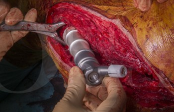
Lernen Sie den totalen Ellenbogenersatz mit dem Coonrad-Morrey Total Elbow Replacement (Zimmer-Biomet) mit Schritt-für-Schritt-Anleitungen auf OrthOracle. Unsere E-Learning-Plattform enthält hochauflösende Bilder und eine zertifizierte CME des chirurgischen Verfahrens für den totalen Ellenbogenersatz mit dem Coonrad-Morrey Total Elbow Replacement (Zimmer-Biomet).
Ein Ellenbogengelenkersatz ist eine attraktive Möglichkeit, Schmerzen bei Patienten mit schmerzhaften arthritischen Gelenken zu lindern, insbesondere im Vergleich zu den Alternativen.
Die ersten Prothesen waren feste Scharniere (die nur Beugung und Streckung zuließen), die aufgrund der normalen Varus- und Valgusbewegungen der Ulna in Bezug auf den Humerus im Gelenk zu vorhersehbarem frühen Lösen der Komponenten führten. Unverbundene Komponenten (bei denen die Humerus- und Unterarmkomponenten nicht physisch verbunden sind und auf das Gleichgewicht der Weichteile für die Stabilität angewiesen sind) wurden entwickelt, aber diese neigten zur Luxation.
Original Intro:
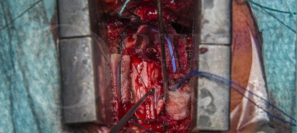
Total elbow replacement using the Coonrad-Morrey Total Elbow Replacement (Zimmer-Biomet)
Learn the Total elbow replacement using the Coonrad-Morrey Total Elbow Replacement (Zimmer-Biomet) surgical technique with step by step instructions on OrthOracle. Our e-learning platform contains high resolution images and a certified CME of the Total elbow replacement using the Coonrad-Morrey Total Elbow Replacement (Zimmer-Biomet) surgical procedure.
An elbow replacement is an attractive proposition to improve pain in patients that have painful arthritic joints, particularly when compared to the alternatives.
Initial prostheses were fixed hinges (allowing only flexion and extension) which, due to the normal varus and valgus toggle of the ulna in relation to the humerus at the joint, led to predictable early loosening of the components. Unlinked components (with the humeral and the forearm components not being physically connected and relying on balancing the soft tissues for stability) were developed, but these were prone to dislocation.
Contemporary implants are therefore usually coupled (physically connected together), but with play in varus and valgus replicating the normal joint to reduce the likelihood of mechanical loosening. Nonetheless, loosening of the components (usually aseptic, but potentially secondary to infection) still frequently occurs, and therefore patients are recommended to restrict activities following elbow replacement for the remainder of their lives, in particular limiting lifting loads and torsion activities. For this reason, the number of patients who will accept the required lifestyle restrictions needed for a replacement to so stand to benefit from elbow replacement after appropriate counselling are relatively limited.
Patients with inflammatory arthritis have generally been the main group of patients who underwent elbow replacement surgery, in large part as their functional demands have tended to be lower due to the involvement of multiple other joints with their disease. With the advent of biologic treatments for systemic inflammatory arthropathies, the number of patients who need to consider having an elbow replacement has reduced. As the number of patients receiving elbow replacements for inflammatory arthritis has reduced, the proportion of patients receiving implantations for un-reconstructible trauma in the elderly, , has increased. The outcomes reported in this group has been good, at least in the medium term. The overall number of primary implantations entered into the UK National Joint Registry each year since data recording began (1st April 2012) has remained around 600 across England and Wales. Most patients having their elbow replaced are still those with an underlying inflammatory arthritis, ideally in their 60’s or older (to minimise the risk of revision surgery being necessary), but if younger patients understand the potential implications of a replacement and accept the restrictions still needed to maximise the longevity of the implant, elbow replacement can be offered to younger patients as well.
The Coonrad-Morrey total elbow replacement is the international market leading elbow replacement implant, used in its current form for around 20 years, with 10-year survivorship of 80-92% reported. In common with most elbow replacements, it is designed for cemented implantation. It represents a reliable, relatively straightforward component to insert with readily available exchange of the polyethylene bearings in the event of significant wear, in the presence of stable implants. The different component sizes have had interchangeable bearings since the early 2000’s to facilitate this.
Newer implants, such as the Latitude EV (Wright Medical), Discovery (Lima) and Nexel (Zimmer-Biomet), are available and are defining their roles in elbow replacement surgery. All have anterior flanges to the humeral component. The Latitude EV can be used as a hemiarthroplasty for treating distal humeral fractures, or as a total elbow replacement, and also has a radial head component. The Latitude also has the option of being inserted coupled (linked)or uncoupled (no linkage between the ulnar and humeral components, which may potentially reduce aseptic loosening), or can even be unlinked at a subsequent procedure once soft-tissue healing has provided stability (or, conversely, be coupled to restore stability if initially inserted unlinked).
The newer implants have advantages in certain technical areas such as easier independent insertion of the ulna and humeral components and where indicated, a radial head component. Whilst the ability to couple the implant once the cemented components are stable may have a theoretical advantages in terms of longevity, none of the newer implants have the data to support a comparable longevity to the Coonrad-Morrey implant. and the surgical techniques and instrumentation are a bit more involved and technically demanding.
I still use the Coonrad-Morrey for most primary replacements, in part due to familiarity, both my own and, as importantly, the theatre staffs. Total elbow replacement even in specialist centres is relatively infrequently performed, the scrub staff will have only a limited exposure to the procedure and the instrumentation. I think under such circumstances it is important to consistently use an appropriate implant, amongst a number of reasons so that the whole team become familiar with the steps and instruments to speed up and improve the operative procedures.
Until more contemporary implants demonstrate a definite survival advantage I will continue to use the Coonrad-Morrey for the majority of my patients, reserving use of a newer implants for younger patients and those in whom I think it is more likely that a revision will become necessary.
Author: Chris Little FRCS (Tr & Orth)
Institution: The Nuffield Orthopaedic centre, Oxford, UK.
zum Artikel auf OrthOracle
zum AOUC-Angebot für OrthOracle