Archiv für den Monat: April 2024

Pilonfraktur: Interne Fixierung unter Verwendung einer Stryker AxSOS 3Ti-Platte

Übersetzt aus dem Englischen:

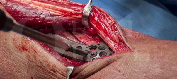
Lernen Sie die chirurgische Technik der Pilonfraktur: Interne Fixierung unter Verwendung einer Stryker AxSOS 3Ti-Platte mit schrittweisen Anweisungen auf OrthOracle. Unsere E-Learning-Plattform enthält hochauflösende Bilder und eine zertifizierte Fortbildung (CME) des chirurgischen Verfahrens der Pilonfraktur: Interne Fixierung unter Verwendung einer Stryker AxSOS 3Ti-Platte.

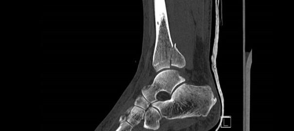
Pilonfrakturen sind Frakturen des distalen Tibiaplafonds und sind per Definition intraartikulär und in einem belasteten Gelenk, was sie zu schweren und oft lebensverändernden Frakturen macht. Die Fraktur entsteht durch eine Mischung aus Scher- und Druckbelastungen auf die distale Tibiametaphyse.

Pilonfrakturen machen zwischen 5 und 10 % aller Frakturen der unteren Extremität aus und sind aufgrund der beteiligten Energie mit einer hohen (15-55 %) Komplikationsrate verbunden. Eine erhebliche Rotationskraft kann auch distale Tibiafrakturen verursachen, die den Plafond einbeziehen, und dies sind ebenfalls Pilonfrakturen. Dieser Mechanismus hat jedoch normalerweise weniger schwere Schädigungen des Weichgewebes und weniger Kompromisse in Bezug auf die Gelenkfläche hinsichtlich Fragmentierung und Knorpelschäden.

Der häufigste Verletzungsmechanismus ist ein Sturz aus großer Höhe, und dieser Fall demonstriert dies, nachdem er durch einen 4 Meter hohen Sturz auf Beton durch ein schwaches Dach verursacht wurde.

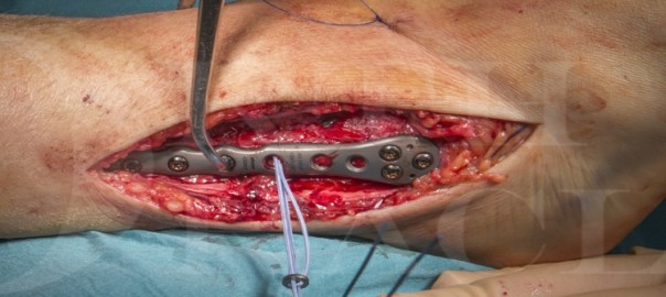
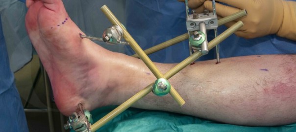
Die große Debatte besteht darin, wann eine offene Repositionsinterne Fixierung (ORIF) oder eine minimalinvasive Plattenosteosynthese (MIPO) verwendet werden soll und wann ein externes Fixationsgestell (wie ein Ilizarov oder ein anderes Drahtkonstrukt) als definitive Behandlung eingesetzt werden soll. In meinen Händen und in einer Einheit mit ausgezeichneten Fertigkeiten in der Drahtfixation vor Ort behandeln wir in der Regel diejenigen Fälle mit sehr schweren Weichteilschäden oder bei denen die Gelenkfläche stark fragmentiert ist, mit einem Gestell. Die Fälle, bei denen die Grad der Gelenkfragmentierung weniger schwerwiegend ist, werden in der Regel mit Plattenfixierung behandelt, wie es bei dem hier vorgestellten Fall der Fall war.

Original Intro:

Pilon fracture: Internal fixation using Stryker AxSOS 3Ti plate

Learn the Pilon fracture: Internal fixation using Stryker AxSOS 3Ti plate surgical technique with step by step instructions on OrthOracle. Our e-learning platform contains high resolution images and a certified CME of the Pilon fracture: Internal fixation using Stryker AxSOS 3Ti plate surgical procedure.

Pilon fractures are fractures of the distal tibial plafond and by definition are intra-articular and in a load bearing joint, this renders them serious and often life changing fractures. The fracture is sustained by a mixture of shear and compressive loads to the distal tibial metaphysis.

Pilon fractures make up between 5 and 10% of all lower limb fractures and because of the energy involved are associated with a high (15 – 55%) complication rate. Significant rotational force can also cause distal tibial fractures which involve the plafond and these are also pilon fractures. This mechanism though usually has less severe soft tissue damage and less compromise to the articular surface in terms of comminution and cartilage damage.

The most frequent mechanism of injury is a fall from height and this case demonstrates this , being sustained after a 4 metre fall onto concrete through a weak roof .

The big debate is when to use ORIF or minimally invasive plate osteosynthesis (MIPO), and when to use an external fixation frame (such as an Ilizarov or other fine wire construct) as definitive treatment. In my hands and working in a unit with excellent fine wire fixation skills locally we tend to treat those cases where there is very severe soft tissue damage or where the articular surface is grossly comminuted with a frame. Those cases where the degree of articular comminution is less severe are usually treated with plate fixation as was the case presented here.

OrthOracle readers will also find the following instructional operative techniques of use:

Pilon fracture: C-type fixed using Smith and Nephew EVOS small fragment system.

Pilon Fracture: C-type fixed with Stryker AxSOS 3 Periarticular Plating System

Pilon fracture: Internal fixation using Stryker AxSOS 3Ti plate.

Ankle fracture: Arthrex tightrope for acute syndesmotic injury and Stryker Variax plate for fibula fracture

Ankle fracture : Fibula pro-tibia fixation technique with Stryker Variax plate.

Ankle fracture: Medial malleolar fixation with ASNIS screws

Ankle fracture: Postero-lateral plating of pronation-external rotation ankle fracture (posterior malleolus))

Ankle fracture: Lateral malleolar fixation using Acumed Fibula Rod System

Author: Mr Chris Blundell FRDS (Tr & Orth)

Institution: IThe Northern General Hospital, Sheffield, UK.

Clinicians should seek clarification on whether any implant demonstrated is licensed for use in their own country.

zum Artikel auf OrthOracle

zum AOUC-Angebot für OrthOracle

Sprunggelenkfraktur: Fixation des hinteren Malleolus unter Verwendung eines posteriomedialen Zugangs

Übersetzt aus dem Englischen:

Lernen Sie die chirurgische Technik der Sprunggelenkfraktur: Fixation des hinteren Malleolus unter Verwendung eines posteriomedialen Zugangs mit schrittweisen Anweisungen auf OrthOracle. Unsere E-Learning-Plattform enthält hochauflösende Bilder und eine zertifizierte Fortbildung (CME) des chirurgischen Verfahrens der Sprunggelenkfraktur: Fixation des hinteren Malleolus unter Verwendung eines posteriomedialen Zugangs.

Sprunggelenkfrakturen mit Beteiligung des hinteren Malleolus gelten als mit einem schlechteren Ergebnis verbunden, jedoch sind das Management und die Indikationen für die Fixierung des hinteren Malleolus noch unzureichend definiert. Die Fixierung von hinteren Malleolusfrakturen wurde erstmals von Lounsbury und Metz im Jahr 1922 beschrieben, die einen extensilen medialen Zugang verwendeten, um große hintere Fragmente mit einem Knochenstift zu fixieren. Die traditionelle Lehre empfahl anschließend die Fixierung von hinteren Malleolusfragmenten basierend auf ihrer Größe, wenn sie auf einem seitlichen Röntgenbild beurteilt wurden, wobei die Fixierung für Fragmente empfohlen wurde, die ein Viertel oder ein Drittel der Gelenkfläche darstellen.

In letzter Zeit hat es einen Paradigmenwechsel in der Behandlung von hinteren Malleolusfrakturen gegeben, da erkannt wurde, dass die Größe des hinteren Malleolusfragments allein nicht der einzige Prädiktor für das Ergebnis ist. Faktoren wie Gelenkkongruenz, hintere Stabilität des Gelenks und Bandstabilität des Sprunggelenks sind ebenfalls wichtige Bestimmer des Ergebnisses.

Original Intro:

Ankle fracture: Posterior malleolar fixation using a posteromedial approach

Learn the Ankle fracture: Posterior malleolar fixation using a posteromedial approach surgical technique with step by step instructions on OrthOracle. Our e-learning platform contains high resolution images and a certified CME of the Ankle fracture: Posterior malleolar fixation using a posteromedial approach surgical procedure.

Ankle fractures with involvement of the posterior malleolus are recognised as having a poorer outcome however the management and indications for fixation of the posterior malleolus remain poorly defined. Fixation of posterior malleolar fractures was first described by Lounsbury and Metz in 1922 who used an extensile medial approach to fix large posterior fragments with a bone peg. Traditional teaching subsequently advocated fixation of posterior malleolar fragments based on their size when assessed on a lateral radiograph with fixation being recommended for fragments representing either a quarter or a third of the articular surface.

Recently there has been a paradigm shift in treatment of posterior malleolar fractures as it has become recognised that the size of the posterior malleolar fragment alone is not the only predictor of outcome. Factors such as articular congruence, posterior stability of the joint and ligamentous stability of the ankle are also important determinants of outcome.

There are a number of classifications describing the posterior malleolus however most are a development of that described by Haraguchi, dividing them into three types based on the appearance on axial CT.  The Haraguchi type 1 injury is a large posterolateral fragment involving the posterior syndesmotic ligament,   a Haraguchi type 2 posterior malleolar fracture extends medially and often has two distinct fragments: posterior and posteromedial with the posteromedial fragment extending to involve the posterior colliculus of the medial malleolus and thus the attachment of the deep deltoid can be rendered unstable by this fragment.  A type 3 injury is a sleeve avulsion of the posterior syndesmotic ligament, this pattern is usually not amenable to fixation and syndesmotic stability is achieved indirectly with syndesmotic screws or equivalent.

Surgical fixation of the posterior malleolus can be performed by a number of approaches.  Traditionally it was achieved with the patient in a supine position, indirect reduction of the fragment and fixation with screws inserted from anterior to posterior to capture the fragment.  It is increasingly recognised however that direct fixation of the fragment via a posterior approach is biomechanically superior.

The most common posterior approach is the posterolateral with an interval between the peronei and flexor hallucis longus, this affords good visualisation of the posterolateral fragment (Haraguchi 1) and also enables fixation of the fibula fracture via the same incision in most cases.  This approach however does not allow fixation of posterior malleolar fractures with medial extension and thus in my hands Haraguchi type 2 fractures are often better fixed via a posteromedial approach.  This can be performed in a variety of ways, often via the bed of tibialis posterior.  In this technique I describe an approach which allows fixation the posteromedial and posterolateral fragments via the same incision utilising windows either side of the posterior neurovascular bundle.

OrthOracle readers will also find the following associated instructional techniques of interest:

Ankle fracture: Postero-lateral plating of pronation-external rotation ankle fracture (posterior malleolus))

Posterior ankle decompression- Open technique

Tarsal tunnel decompression (for tarsal tunnel syndrome)

Ankle fracture: Medial malleolar fixation with ASNIS screws

Ankle fracture : Fibula pro-tibia fixation technique with Stryker Variax plate.

Ankle fracture: Lateral malleolar fixation using Acumed Fibula Rod System

Ankle fracture: Arthrex tightrope for acute syndesmotic injury and Stryker Variax plate for fibula fracture

Author: Paul Fenton, FRCS (Tr & Orth)

Institution: The Queen Elisabeth Hospital, Birmingham, UK.

Clinicians should seek clarification on whether any implant demonstrated is licensed for use in their own country.

zum Artikel auf OrthOracle

zum AOUC-Angebot für OrthOracle

Sprunggelenkfraktur: Arthrex TightRope für akute Syndesmosenverletzung und Stryker Variax-Platte für Fibulafraktur

Übersetzt aus dem Englischen:

Lernen Sie die chirurgische Technik der Sprunggelenkfraktur: Arthrex TightRope für akute Syndesmosenverletzung und Stryker Variax-Platte für Fibulafraktur mit schrittweisen Anweisungen auf OrthOracle. Unsere E-Learning-Plattform enthält hochauflösende Bilder und eine zertifizierte Fortbildung (CME) des chirurgischen Verfahrens der Sprunggelenkfraktur: Arthrex TightRope für akute Syndesmosenverletzung und Stryker Variax-Platte für Fibulafraktur.

Die distale Tibiofibulare Syndesmose ist ein einzigartiges syndesmotisches Gelenk, das durch eine Vielzahl von Bändern kraftvoll verbunden ist, um die Integrität des Sprunggelenks zu erhalten. Die distale Anatomie der beiden Knochen ist so gestaltet, dass die mediale Oberfläche der distalen Fibula in eine Rille auf der lateralen Oberfläche der distalen Tibia passt, die als Incisura fibularis bezeichnet wird und diesem Gelenk die knöcherne Stabilität bietet. Der vordere Teil dieses Gelenks wird durch das vordere untere distale Tibiofibulare Band (AITFL), der hintere Aspekt durch das hintere untere distale Tibiofibulare Band (PITFL) und das transversale Tibiofibulare Band stabilisiert. Direkt zwischen den benachbarten Oberflächen der Tibia und Fibula befindet sich das interosseöse Band, das sich über die gesamte Länge der Fibula erstreckt.

Das PITFL ist bei weitem das stärkste Band in diesem Komplex und am wenigsten wahrscheinlich zu reißen. Wenn es jedoch einmal gerissen ist, ist es am wahrscheinlichsten mit schweren Rotations- oder Luxationsverletzungen des Sprunggelenks und damit verbundener Sprunggelenkinstabilität verbunden.

Original Intro:

Ankle fracture: Arthrex tightrope for acute syndesmotic injury and Stryker Variax plate for fibula fracture

Learn the Ankle fracture: Arthrex tightrope for acute syndesmotic injury and Stryker Variax plate for fibula fracture surgical technique with step by step instructions on OrthOracle. Our e-learning platform contains high resolution images and a certified CME of the Ankle fracture: Arthrex tightrope for acute syndesmotic injury and Stryker Variax plate for fibula fracture surgical procedure.

The distal tibiofibular syndesmosis is a unique syndesmotic joint, powerfully bound by a variety of ligaments to maintain the integrity of the ankle mortise. The distal anatomy of the two bones are created in such a way that the medial surface of the distal fibula fits into a groove on the lateral surface of the distal tibia called the incisura fibularis which offers the bony stability to this joint. The anterior part of this joint is stabilised by the Anterior Inferior Distal Tibio-Fibular ligament (AITFL), the posterior aspect by the Posterior Inferior Distal Tibio-Fibular ligament (PITFL) and the transverse Tibio-Fibular ligament. Directly between the contiguous surfaces of the tibia and fibula is the interosseous ligament which extends throughout the length of the fibula.

The PITFL is by far the strongest ligament in this complex and is the least likely to be ruptured. Once ruptured however it is most likely to be associated with severe rotational or dislocating injuries of the ankle and associated ankle instability.

The syndesmotic ligaments  stabilise the fibula with respect to both talus and tibia, whilst allowing a degree of rotation of the fibula, required in particular during full ankle dorsiflexion when the widest portion of the talus comes into the mortise. As with any ligament injury the key point is not solely whether these ligaments have been injured, but rather their stability in determining the need for treatment. If instability is not addressed, high contact pressures at the joints surface ensue and early degenerative change occurs in most patients.

Traditional fixation of unstable syndesmotic injuries has been with two parallel, non-compressive, small fragment screws placed across the ankle at the level of the tibial incisura. Such fixation also requires a decision as to whether the screws need to be removed and furthermore,  the optimal timing for such removal.

The use of a Arthrex tightrope implant, is far more physiological in terms of the way the syndesmosis is held, allowing more normal movement at the ankle mortise. It is a simple and ingenious implant that has proved its worth not only in syndesmotic ankle reconstruction but also in the shoulder and forefoot. It consists of two metal buttons that rest on the respective bony  surfaces to be approximated, and a robust four stranded Fibrewire suture construct which links these buttons. The design allows easy apposition of the buttons towards each other by the effective “slip-knot” set up of the suture strands.

Readers will also find of use the following OrthOracle techniques:

Internal fixation of ankle fracture : Fibula pro-tibia fixation technique with Stryker Variax plate.

Internal fixation of medial malleolar ankle fracture with ASNIS screws

Lateral malleolar fixation using Acumed Fibula Rod System

Postero-lateral plating of pronation-external rotation ankle fracture (posterior malleolar fixation)

Fusion of the syndesmosis for isolated distal tibio-fibular arthritis

Author: Mark Herron FRCS

Institution: The Wellington Hospital, London, UK.

Clinicians should seek clarification on whether any implant demonstrated is licensed for use in their own country.

zum Artikel auf OrthOracle

zum AOUC-Angebot für OrthOracle

Sprunggelenkfraktur: Stabilisierung mit Hoffmann 3 Sprunggelenk-übergreifendem externem Fixateur (Delta-Rahmen)

Übersetzt aus dem Englischen:

Lernen Sie die chirurgische Technik der Sprunggelenkfraktur: Stabilisierung mit Hoffmann 3 Sprunggelenk-übergreifendem externem Fixateur (Delta-Rahmen) mit schrittweisen Anweisungen auf OrthOracle. Unsere E-Learning-Plattform enthält hochauflösende Bilder und eine zertifizierte Fortbildung (CME) des chirurgischen Verfahrens der Sprunggelenkfraktur: Stabilisierung mit Hoffmann 3 Sprunggelenk-übergreifendem externem Fixateur (Delta-Rahmen).

Die externe Fixation des Sprunggelenks ist ein wesentlicher Eingriff, der für jeden Chirurgen, der akute Traumafälle behandelt, zur Routine werden muss. Obwohl Sprunggelenkfrakturen häufig sind, müssen die meisten nicht mit einem temporären übergreifenden externen Fixateur behandelt werden, da es sich in der Regel um geschlossene Verletzungen niedriger Energie handelt. Sprunggelenk-übergreifende externe Fixateure sind angezeigt bei Verletzungen höherer Energie, bei denen eine “Überbrückung, Untersuchung und Planung” erforderlich ist, d. h. bei einer Pilonfraktur; einer offenen Sprunggelenkfraktur; einer Talusdislokation oder bei Verletzungen mit signifikant traumatisiertem Weichgewebe. Dennoch sollte der erste Schritt in Ihrem Behandlungsalgorithmus eine sofortige Gelenk- / Frakturreduktion und die Anwendung eines Gipsverbandes sein. Bei grob instabilen Verletzungen, solchen, die nicht reduziert werden können, solchen, bei denen die Frakturlänge nicht aufrechterhalten werden kann, oder solchen, die eine Überwachung des Weichgewebes erfordern, ist ein übergreifender externer Fixateur eine bessere Option.

Der Standard des Britischen Orthopädischen Verbandes für Traumata (BOAST) empfiehlt bei instabilen Sprunggelenkfrakturen in der Altersgruppe unter 60 Jahren eine frühzeitige operative Fixierung (d. h. <24 Stunden). Bei der Altersgruppe über 60 Jahren kann, sofern eine zufriedenstellende Reduktion erreicht und aufrechterhalten werden kann, eine enge Kontaktgipsschiene eine Option sein. Die Richtlinien besagen, dass die Verwendung einer externen Fixation bei grober Instabilität in Verbindung mit einer Beeinträchtigung des Weichgewebes selten indiziert sein kann. Dies impliziert daher, dass Patienten mit einer Fraktur-Dislokation, die älter als 24 Stunden ist, nicht reduziert und in einem Gipsverband aufrechterhalten werden kann, für einen übergreifenden Sprunggelenk-externen Fixateur in Betracht gezogen werden sollten. Meiner Erfahrung nach können Patienten zwischen 24 und 48 Stunden eine sich entwickelnde Verletzung haben, und die Schwellung kann noch zunehmen. Dies bedeutet, dass bei Anwendung eines Gipsverbandes dieser aufgrund anhaltender Schwellung gelöst werden muss und die Frakturreduktion verloren geht.

mehr

Original Intro:

Ankle fracture: Stabilisation with Hoffmann 3 Ankle-spanning External Fixator (Delta frame)

by Ross Fawdington FRCS (Tr & Orth)

Learn the Ankle fracture: Stabilisation with Hoffmann 3 Ankle-spanning External Fixator (Delta frame) surgical technique with step by step instructions on OrthOracle. Our e-learning platform contains high resolution images and a certified CME of the Ankle fracture: Stabilisation with Hoffmann 3 Ankle-spanning External Fixator (Delta frame) surgical procedure.

Ankle external fixation is an essential procedure that needs to be second nature for any surgeon undertaking acute trauma cases. Although ankle fractures are common, the vast majority do not need to be treated with a temporary spanning external fixator because they are usually low energy, closed injuries. Ankle spanning external fixators are indicated for higher energy injuries where there is a need to “span, scan and plan” i.e. a pilon fracture; an open ankle fracture; a talar body dislocation, or those where there are significantly traumatised soft tissues. Having said that, the first step in your treatment algorithm should be immediate joint / fracture reduction and application of a plaster of Paris backslab. In grossly unstable injuries, those that cannot be reduced, those where fracture length cannot be maintained or those that require soft tissue monitoring, then a spanning external fixator is a better option.

The British Orthopaedic Association Standard for Trauma (BOAST) recommends for unstable ankle fractures in the under 60 age group early operative fixation (i.e. < 24 hours). In the over 60 age group,  providing a satisfactory reduction can be achieved and maintained, then a close contact cast is an option. The guidelines state that the use of external fixation may be rarely indicated in the presence of gross instability associated with soft tissue compromise. This therefore implies that patient’s with a fracture dislocation, older than 24 hours that cannot be reduced and maintained in a plaster of Paris, should be considered for a spanning ankle external fixator. It is my experience, that patients between 24-48 hours can have an evolving injury and the swelling may still be on the way up. This means that if a plaster backslab is applied, it may need to be released due to ongoing swelling and the fracture reduction is lost.

There are also a cohort of patients that as the swelling decreases, the plaster backslab becomes loose and the fracture re-displaces and thus the swelling persists. There’s a final group that due to repeated soft tissue inspection, the plaster backslab becomes weaker / looser and again the fracture re-displaces and the swelling persists. In general, if a patient still has significant swelling at day 5 post reduction, then I’d recommend checking with an x-ray, as occasionally the fracture has subluxed and this can also happen while in an external fixator depending on the stability of the construct.

The Hoffmann 3 external fixation system was developed for use in acute trauma, “damage control” orthopaedics and also for definitive fixation. It has a variety of features that make it simple, fast to apply and adaptable for many types of patients and injury patterns. It is indicated for stabilisation of open and/or unstable fractures and where soft tissue injury may preclude the use of other fracture treatments e.g. casts, intramedullary devices or internal fixation. Additional indications include:

  • Bone fracture fixation
  • Osteotomy
  • Arthrodesis
  • Correction of deformity
  • Revision procedure e.g. infection
  • Bone reconstruction procedures

The Hoffmann 3 system is MRI Conditional up to 3.0 Tesla. This means that patient’s with this type of fixator can go into an MRI scanner providing certain conditions are met e.g. the field strength does not exceed 3.0 Tesla, and the MRI scan is performed in accordance with the testing schedule as specified by the manufacturer. The Delta couplings are fully compatible with the Hoffmann II MRI and the Hoffmann II Compact MRI. This means that you can use any combination of 5mm, 8mm and 11mm connecting rods.

OrthOracle readers will also find the following associated operative techniques of interest:

Minimally invasive distal tibial osteotomy and correction of deformity with the Taylor Spatial Frame

Compartment fasciotomy and Hoffmann 3 spanning external fixator for open tibial fracture

Tibial fracture non-union correction using Taylor Spatial Frame (Smith and Nephew)

Tibial shaft fracture: Fixation with a Taylor Spatial Frame (TSF) circular external fixator (Smith and Nephew)

Distal radius fracture: Compound injury stabilised with Hoffman II External Fixator

Author: Ross Fawdington FRCS (Tr & Orth)

Institution: The Queen Elisabeth Hospital, Birmingham, UK.

Clinicians should seek clarification on whether any implant demonstrated is licensed for use in their own country.

zum AOUC-Angebot für OrthOracle

Let’s discuss Osteo: Pseudarthrose

Die Behandlung von Pseudarthrosen bleibt eine Herausforderung in der Unfallchirurgie und Orthopädie. Die operative Behandlung richtet sich nach der betroffenen Körperregion sowie der Schwere des Traumas. Dabei gilt: Je komplexer die Fraktur, desto anspruchsvoller ist die Versorgung, denn es gibt viel zu beachten! Im Vortrag „Pseudarthrose und chirurgische Intervention“ erfahren Sie von Univ.-Prof. Dr. med. Uwe Maus, Düsseldorf, welche chirurgischen Maßnahmen in welchen Fällen sinnvoll sind. Das Online-Seminar ist Teil der beliebten Reihe „Let’s discuss Osteo“ auf dem Fortbildungsportal www.Facts4Fractures.de.

Online-Seminar mit Univ.-Prof. Dr. med. Uwe Maus

Wie kann Ihr genauer Blick dazu beitragen, Pseudarthrosen vollumfänglich abzuklären und die optimale chirurgische Intervention zu wählen? Das aktuelle Seminar der erfolgreichen Fortbildungsreihe ist nun on demand verfügbar:

Referent: Univ.-Prof. Dr. med. Uwe Maus, Düsseldorf
Moderator:
Prof. Dr. med. Christopher Niedhart, Heinsberg
Veranstalter: med-kompakt, unterstützt von Alexion

Das nächste Online-Seminar der Reihe „Let’s discuss Osteo“ ist bereits in Planung:

09.10.2024

Kraniosynostosen – Ursachen und therapeutische Optionen


Prof. Dr. med. Tilmann Schweitzer

Jetzt anmelden

Sie möchten kein Seminar verpassen? Dann lassen Sie sich unkompliziert und bequem per Mail erinnern und melden Sie sich für den Info-Service an.

Falls Sie nicht live dabei sein können, sind alle Seminare im Nachgang on demand verfügbar. Zuletzt sprach PD Dr. med. Lothar Seefried über „Seltene Knochenerkrankungen im Praxisalltag“: www.Facts4Fractures.de

Differentialdiagnose Hypophosphatasie

Die chirurgische Intervention ist ein wichtiger Baustein bei der Behandlung von Pseudarthrosen. Doch dafür ist es wichtig zu verstehen, weshalb die Fraktur nicht verheilt ist. Mögliche Ursachen sind beispielsweise eine unzureichende Stabilisierung oder zu frühe Belastung des Bruchs, mangelnde Durchblutung oder Infektionen.1 Doch es gibt eine wichtige, oft übersehene Differentialdiagnose: Hypophosphatasie (HPP). Diese fortschreitende und seltene Knochenmineralisierungsstörung kann schwere Auswirkungen auf verschiedene Körperfunktionen haben.2,3,4 Durch einen Mangel an alkalischer Phosphatase kommt es bei den Patient:innen zur Osteomalazie, welche wiederum Knochenbrüche und Pseudarthrose begünstigen kann. Ein Warnsignal ist eine erniedrigte alkalische Phosphatase.2,3,4,5,6,7 

Erfahren Sie kurz und bündig mehr zu Diagnostik und klinischem Erscheinungsbild von HPP.


Die Diagnose seltener Erkrankungen ist oft ein langer Weg. In der aktuellen Episode von „Let’s talk Osteo“ diskutiert ein Arzt mit seiner HPP-Patientin darüber, wie sie diesen erlebten. Was waren die ersten Anzeichen? Wo wurde falsch abgebogen? Und was führte schließlich zum Ziel? Das bespricht Prof. Dr. med. Dr. h.c. Christian Wüster mit seiner Patientin, die erst nach vielen Umwegen die korrekte Diagnose HPP erhielt. Freuen Sie sich auf eine inspirierende und informative Diskussion!


Forum Knochenstoffwechsel: Jetzt herunterladen

Welche Laborwerte sollten Sie bei Knochenstoffwechselerkrankungen besonders beachten? Und wie sind diese zu deuten? In der aktuellen Ausgabe des „Forum Knochenstoffwechsel“ finden Sie die wesentlichen Informationen kompakt aufbereitet. Erfahren Sie außerdem, welchen Einfluss Bewegung und Muskelaktivität auf die Knochengesundheit hat und wie gezielte Fördermaßnahmen das Muskel-Knochen-Zusammenspiel verbessern. Hier können Sie auch vom wohl berühmtesten Bergsteiger „Ötzi“ lernen. Holen Sie sich jetzt das e.PDF.

_____________________________________________________________

Quellenverzeichnis:

1 Rüter A et al., Chirurg. 1999; 70:1239-1245.

2 Briot K et al., Arch Pediatr. 2017; 24(5S2):5S71-5S73.

3 Bianchi ML et al., Osteoporosis International. 2020; 31:1445-1460.

4 Beck C et al., Open Bone J. 2009; 1:8-15.

5 Weber TJ et al., Metabolism. 2016; 65:1522-1530.

6 Högler W et al., BMC Musculoskelet Disord. 2019; 20:80.

7 Seefried L et al., JBMR. 2020; 35:2171-2178.